. 2012 Jun 15;3(1-3):121-126.
doi: 10.3727/215517912X639441. eCollection 2012 Jan.

Isolation of Hepatic Progenitor Cells From Human Liver With Cirrhosis Secondary to Biliary Atresia Using EpCAM or Thy-1 Markers

Affiliations

Isolation of Hepatic Progenitor Cells From Human Liver With Cirrhosis Secondary to Biliary Atresia Using EpCAM or Thy-1 Markers

Taisuke Yamazaki et al. Cell Med. .

Abstract

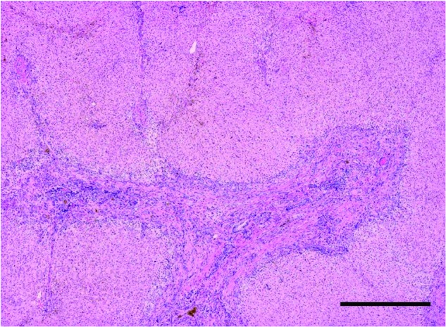
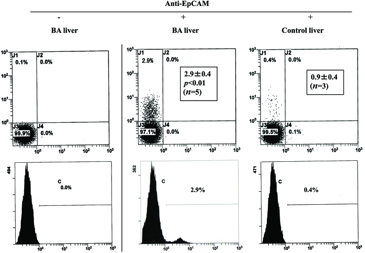
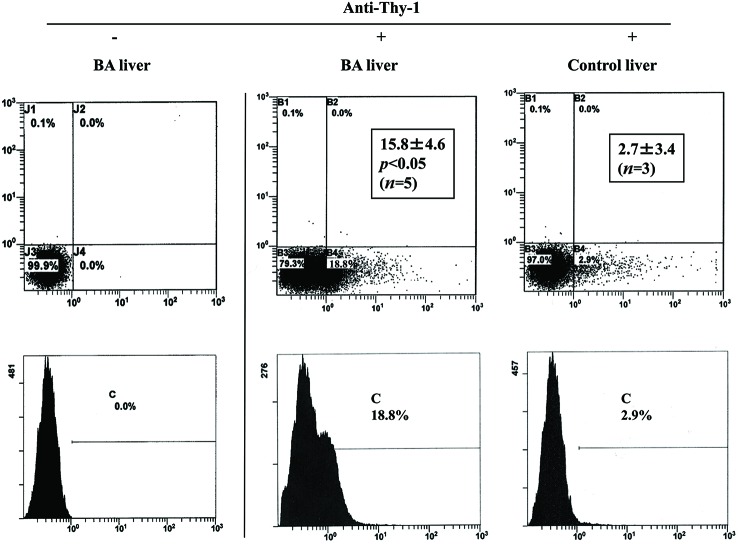
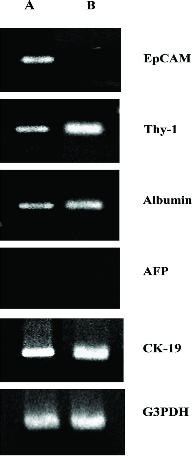
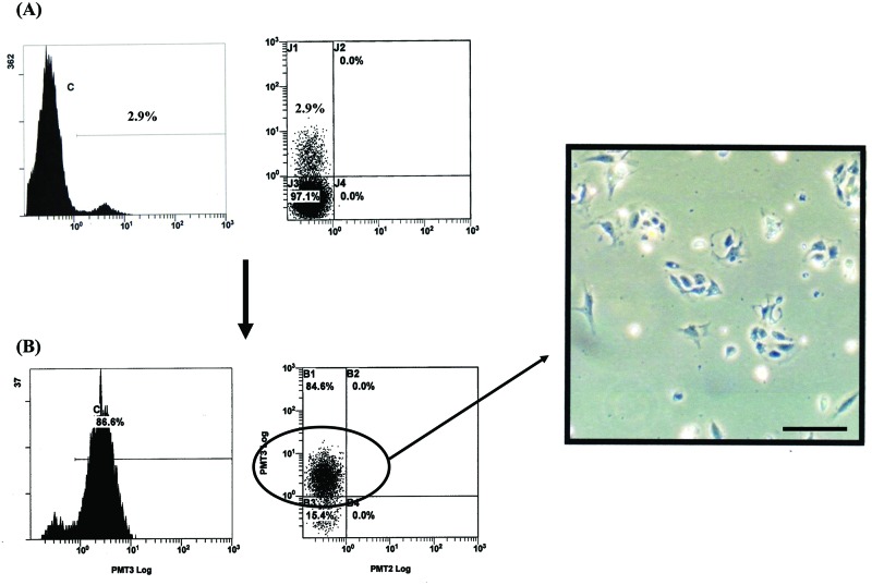
We sought to determine whether hepatic progenitor cells can be isolated from cirrhotic liver using epithelial cell adhesion molecule (EpCAM) or Thy-1 markers. Liver tissue with cirrhosis secondary to biliary atresia (BA) was collagenase digested, and nonparenchymal cells (NPCs) were cultivated for 24 h. Noncirrhotic NPCs derived from patients with carbamyl phosphate synthetase and ornithine transcarbamylase deficiencies were used as controls. Flow cytometric analysis demonstrated that the percentages of EpCAM- and Thy-1-positive cells were significantly higher in NPC populations derived from BA liver than in those derived from control liver. Reverse transcription polymerase chain reaction analysis revealed that EpCAM-positive sorted cells expressed EpCAM, Thy-1, albumin, and CK-19, whereas Thy-1-positive sorted cells expressed Thy-1, albumin, and CK-19. These findings indicate that EpCAM- or Thy-1-positive hepatic progenitor cells can be more efficiently isolated from BA liver than from control liver and suggest that the properties of EpCAM-positive cells are somewhat different from those of Thy-1-positive cells.

Keywords: Biliary atresia; Cirrhosis; EpCAM; Flow cytometry; Hepatic progenitor cells; Thy-1.


Figures

Figure 1
Figure 1
Histology of liver tissue from a patient with cirrhosis secondary to biliary atresia (BA). Hematoxylin and eosin staining. Scale bar: 250 μm.
Figure 2
Figure 2
Flow cytometric analysis of nonparenchymal cell (NPC) populations derived from biliary atresia (BA) and control liver. Cells were stained with (+) or without (−) phycoerythrin (PE)-labeled anti-epithelial cell adhesion molecule (EpCAM) antibody and were analyzed using flow cytometry. Insets indicate the mean (±SD) percentage of EpCAM-positive cells.
Figure 3
Figure 3
Flow cytometric analysis of nonparenchymal cell (NPC) populations derived from biliary atresia (BA) and control liver. Cells were stained with (+) or without (−) fluorescein isothiocyanate-labeled anti-mouse monoclonal Thy-1 antibody and were analyzed using flow cytometry. Insets indicate mean (±SD) percentage of Thy-1-positive cells.
Figure 4
Figure 4
Reverse transcription and polymerase chain reaction (RT-PCR) analysis of epithelial cell adhesion molecule (EpCAM)- and Thy-1-positive sorted cells derived from biliary atresia (BA) liver. The indicated mRNA expression was assessed using RT-PCR and (A) EpCAM-positive cells (A) or Thy-1- positive cells (B). G3PDH was included as an internal loading control. The results are representative of two independent experiments. AFP, α-fetoprotein; G3PDH, glyceraldehyde-3-phosphate dehydrogenase.
Figure 5
Figure 5
Culture of epithelial cell adhesion molecule (EpCAM)-positive sorted cells derived from biliary atresia (BA) liver. Flow cytometric analysis of cells from BA liver before (A) and after (B) cell sorting for EpCAM-positive cells. The cells in (B) were epithelial-like on day 2 of culture as determined by phase contrast microscopy. Scale bar: 250 μm.

References

    1. Crosby H. A.; Kelly D. A.; Strain A. J. Human hepatic stemlike cells isolated using c-kit or CD34 can differentiate into biliary epithelium. Gastroenterology 120:534–544;2001. - PubMed
    1. Dezso K.; Jelnes P.; Laszlo V.; Baghy K.; Bodor C.; Paku S.; Tygstrup N.; Bisgaard H. C.; Nagy P. Thy-1 is expressed in hepatic myofibroblasts and not oval cells in stem cell-mediated liver generation. Am. J. Pathol. 171:1529–1537;2007. - PMC - PubMed
    1. Enosawa S. Hepatocyte isolation from human liver tissue. Organ Biol. 16:361–370;2009.
    1. Herrera M. B.; Bruno S.; Buttiglieri S.; Tetta C.; Gatti S.; Deregibus M. C.; Bussolati B.; Camussi G. Isolation and characterization of a stem cell population from adult human liver. Stem Cells 24:2840–2850;2006. - PubMed
    1. Hussain S. Z.; Strom S. C.; Kirby M. R.; Burns S.; Langemeijer S.; Ueda T.; Hsieh M.; Tisdale J. F. Side population cells derived from adult human liver generate hepatocytelike cells in vitro. Dig. Dis. Sci. 50:1755–1763;2005. - PMC - PubMed