. 2017 Mar;47(3):458-469.
doi: 10.1002/eji.201646855. Epub 2017 Jan 25.

Deletional tolerance prevents AQP4-directed autoimmunity in mice

Affiliations

Deletional tolerance prevents AQP4-directed autoimmunity in mice

Anna-Lena Vogel et al. Eur J Immunol. 2017 Mar.

Abstract

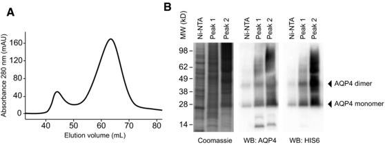
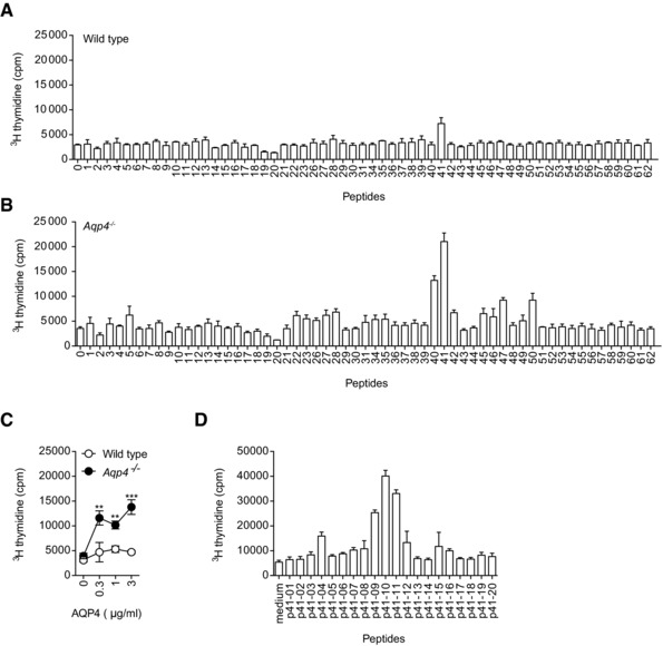
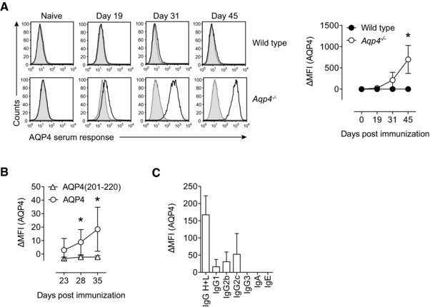
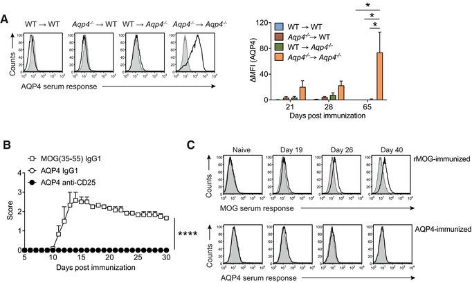
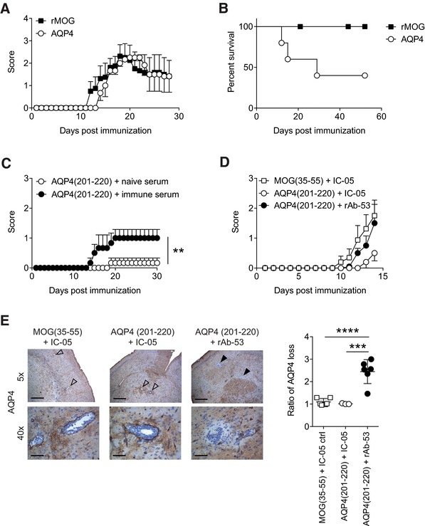
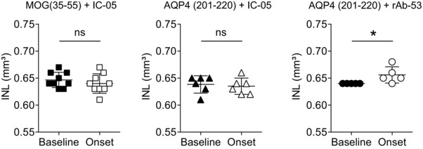
Neuromyelitis optica (NMO) is an autoimmune disorder of the central nervous system (CNS) mediated by antibodies to the water channel protein AQP4 expressed in astrocytes. The contribution of AQP4-specific T cells to the class switch recombination of pathogenic AQP4-specific antibodies and the inflammation of the blood-brain barrier is incompletely understood, as immunogenic naturally processed T-cell epitopes of AQP4 are unknown. By immunizing Aqp4-/- mice with full-length murine AQP4 protein followed by recall with overlapping peptides, we here identify AQP4(201-220) as the major immunogenic IAb -restricted epitope of AQP4. We show that WT mice do not harbor AQP4(201-220)-specific T-cell clones in their natural repertoire due to deletional tolerance. However, immunization with AQP4(201-220) of Rag1-/- mice reconstituted with the mature T-cell repertoire of Aqp4-/- mice elicits an encephalomyelitic syndrome. Similarly to the T-cell repertoire, the B-cell repertoire of WT mice is "purged" of AQP4-specific B cells, and robust serum responses to AQP4 are only mounted in Aqp4-/- mice. While AQP4(201-220)-specific T cells alone induce encephalomyelitis, NMO-specific lesional patterns in the CNS and the retina only occur in the additional presence of anti-AQP4 antibodies. Thus, failure of deletional T-cell and B-cell tolerance against AQP4 is a prerequisite for clinically manifest NMO.

Keywords: Aquaporin 4 (AQP4); B cell; Encephalitogenic epitope; Experimental neuromyelitis optica; Optical coherence tomography; T cell; Tolerance.


Figures

Figure 1
Figure 1
Protein expression and purification. The full‐length AQP4 isoform M1 was produced in High Five cells using the Baculovirus‐insect cell expression system. (A) Purification of the protein by ultracentrifugation, Ni‐NTA affinity chromatography, and subsequent gel filtration on a S200 HR 16/60 column. n‐Octyl‐β‐d‐glucopyranoside was used as detergent throughout the process. (B) Side‐by‐side analysis of the Ni‐NTA elution fraction as well as the pooled fractions of gel filtration peaks 1 and 2 using Coomassie staining and western blot analysis for detection of AQP4 and HIS6. Data are representative of six independent experiments (A, B).
Figure 2
Figure 2
AQP4(201–220) is the immunodominant IAb‐restricted epitope of AQP4. WT C57BL/6 and Aqp4 −/− mice were immunized s.c. with full‐length AQP4 protein (A, B, and D) or AQP4(201–220) peptide (C) emulsified in CFA. Draining LN cells and splenocytes were analyzed for cell proliferation in response to single peptides or full‐length AQP4 protein. (A, B) Splenocytes and LN cells from AQP4 immunized WT and Aqp4 −/− mice were stimulated with single 20‐mer peptides spanning the whole sequence of AQP4. (C) Draining LN cells of AQP4(201–220) immunized WT and Aqp4 −/− mice were tested for their proliferative recall response to full‐length AQP4 protein. (D) Fine mapping of the immunodominant AQP4 epitope was performed using 11‐mer peptides spanning AQP4(196–225) to recall cells from AQP4‐immunized Aqp4 −/− mice. Proliferation was measured by 3[H]‐thymidine incorporation. Means of triplicate cultures ± SD are shown. **p < 0.01, ***p < 0.001 (Student's t‐test). Data are representative of five independent experiments (A–D).
Figure 3
Figure 3
Aqp4 −/− mice but not WT controls mount a robust antibody response to AQP4 upon immunization with full‐length AQP4 protein. WT C57BL/6 and Aqp4 −/− mice were immunized s.c. with full‐length AQP4 protein or AQP4(201–220) peptide emulsified in CFA. (A) Sera of naive mice and AQP4‐immunized WT or Aqp4 −/− mice at different time points after immunization with AQP4 were analyzed for AQP4‐specific antibodies in a cell‐based flow cytometric assay with LN18 cells transduced with AQP4 expressing lentivirus. Anti‐mouse total IgG H+L (AlexaFluor488 labeled) was used to detect anti‐AQP4 antibodies (black line histograms). The ΔMFI was calculated in relation to staining of LN18 cells transduced with empty vector (shaded histograms). Representative histograms (left) and plot of ΔMFI (right). Mean ΔMFI ± SD (n = 6 per group). (B) AQP4 serum response at different time points in Aqp4 −/− mice that were immunized with AQP4 protein or AQP4 (201–220) peptide. Mean ΔMFI ± SD (n = 6 per group). (C) To specify the antibody classes and subclasses, fluorochrome‐labeled anti‐mouse Ig antibodies specific for IgA, IgE, IgG1, IgG2b, IgG2c, and IgG3 were used. Mean ΔMFI ± SD (n = 6). *p < 0.05 (unpaired Student's t‐test). Data are representative of three independent experiments (AC).
Figure 4
Figure 4
The naive T‐cell and B‐cell repertoires of WT mice are purged of AQP4 reactive clones. (A) Criss‐cross BM chimeras of WT C57BL/6 and Aqp4 −/− mice were immunized with full‐length AQP4 protein emulsified in CFA. Sera from different time points after immunization were tested for AQP4‐specific antibodies in a cell‐based flow cytometric assay as described in Fig. 3. Representative histograms (left) and Mean ΔMFI ± SD (n = 6 per group) for AQP4‐specific IgG (right). *p < 0.05 (ANOVA plus Sidak's post‐test). (B) WT C57BL/6 mice were immunized s.c. with full‐length AQP4 protein or MOG(35–55) emulsified in CFA as immunization control. On days –5 and –3 prior to immunization, some mice were injected i.p. with control IgG1 or with anti‐CD25 antibodies to deplete Treg cells before immunization with full‐length AQP4 protein. Mean clinical scores ± SEM (n = 6 per group). ****p < 0.0001 (Mann–Whitney U test for nonparametric values). (C) Aqp4 ΔT x Tcra −/− mice were generated by i.p. transfer of the mature T‐cell compartment of Aqp4 −/− mice into Tcra −/− mice. The mice were immunized with full‐length rMOG or AQP4 protein emulsified in CFA. Sera of mice from each group were collected at different time points after immunization and tested for MOG‐ and AQP4‐specific antibodies in a cell‐based flow cytometric assay as described. Representative histogram plots illustrating the anti‐MOG and anti‐AQP4 serum responses in Aqp4 ΔT × Tcra −/− mice at different time points (n = 6 per group). Data are representative of two independent experiments (C).
Figure 5
Figure 5
AQP4(201–220) is an encephalitogenic epitope. (A, B) Aqp4 ΔT × Tcra −/– mice were immunized with full‐length AQP4 or rMOG protein emulsified in CFA and monitored for symptoms of encephalomyelitis. (A) Mean clinical scores ± SEM and (B) percent survival of immunized mice are shown (n = 6 per group). (C) Aqp4 ΔT × Rag1 −/− mice were immunized with AQP4(201–220) peptide emulsified in CFA. One hundred microliters of naïve serum or immune serum with high titers of anti‐AQP4 antibodies (harvested from AQP4‐immunized Aqp4 −/− mice) were administered i.v. on days 6 and 12 after immunization. Mean clinical scores ± SEM (n = 5 per group). **p < 0.01 (Mann–Whitney U test for nonparametric values). (D, E) Aqp4 ΔT × Rag1 −/− mice were immunized with either AQP4(201–220) or MOG(35–55) peptide emulsified in CFA. On days 12 and 14 after immunization, the mice were injected i.v. with rAb‐53 antibody or IC‐05 control antibody. (D) Mean clinical scores ± SEM (n = 6 per group). (E) Mice were sacrificed 4 h after the last antibody treatment to perform histological analysis. Representative AQP4 staining of the brain at 5× (scale bar, 400 μm) and 40× magnification (scale bar, 50 μm). Open arrows show vessels without perivascular loss of AQP4 immunoreactivity. Closed arrows indicate areas of AQP4 loss in the vicinity of vessels. Quantification of AQP4 loss as ratio of the area with AQP4 signal loss divided by the area of the associated vessel lumen in the brain of the indicated treatment groups (mean ± SD). ***p < 0.001; ****p < 0.0001 (ANOVA plus Sidak's post‐test). Data are representative of two independent experiments (A, B, D, and E).
Figure 6
Figure 6
Aqp4 ΔT × Rag1 −/− mice immunized with AQP4(201–220) peptide plus i.v. application of rAb‐53 develop inner nuclear layer (INL) swelling at disease onset. Aqp4 ΔT × Rag1 −/− mice were immunized with either AQP4(201–220) or MOG(35–55) peptide emulsified in CFA. After immunization, the mice were injected i.v. with rAb‐53 antibody or IC‐05 control antibody on day 12 after immunization. Retinal optical coherence tomography (OCT) was performed in the indicated treatment groups prior to immunization (baseline) and on day 13 after immunization at the onset of disease (and 1 day after administration of control antibody IC‐05 or anti‐AQP4 antibody rAb‐53). Mean INL volumes of individual eyes ± SD are shown. *p < 0.05, ns = not significant (Student's t‐test). One experiment with six mice per group is shown.

References

    1. Papadopoulos, M. C. and Verkman, A. , Aquaporin 4 and neuromyelitis optica. Lancet Neurol. 2012. 11: 535–544. - PMC - PubMed
    1. Verkman, A. S. , Aquaporins in clinical medicine. Annu. Rev. Med. 2012. 63: 303–316. - PMC - PubMed
    1. Delarasse, C. , Daubas, P. , Mars, L. T. , Vizler, C. , Litzenburger, T. , Iglesias, A. , Bauer, J. et al., Myelin/oligodendrocyte glycoprotein‐deficient (MOG‐deficient) mice reveal lack of immune tolerance to MOG in wild‐type mice. J. Clin. Invest. 2003. 112: 544–553. - PMC - PubMed
    1. Lennon, V. A. , Wingerchuk, D. M. , Kryzer, T. J. , Pittock, S. J. , Lucchinetti, C. F. , Fujihara, K. , Nakashima, I. et al., A serum autoantibody marker of neuromyelitis optica: distinction from multiple sclerosis. Lancet 2004. 364: 2106–2112. - PubMed
    1. Lucchinetti, C. F. , Mandler, R. N. , McGavern, D. , Bruck, W. , Gleich, G. , Ransohoff, R. M. , Trebst, C. et al., A role for humoral mechanisms in the pathogenesis of Devic's neuromyelitis optica. Brain. 2002. 125: 1450–1461. - PMC - PubMed

Publication types

MeSH terms

LinkOut - more resources