. 2017 Sep;37(9):3203-3218.
doi: 10.1177/0271678X16685106. Epub 2017 Jan 6.

Intranasal insulin treatment of an experimental model of moderate traumatic brain injury

Affiliations

Intranasal insulin treatment of an experimental model of moderate traumatic brain injury

Fiona Brabazon et al. J Cereb Blood Flow Metab. 2017 Sep.

Abstract

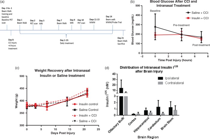
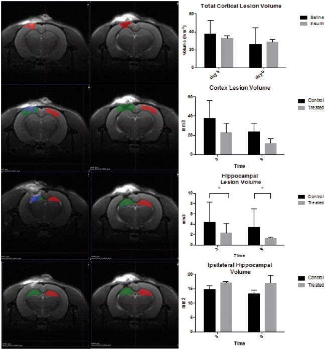
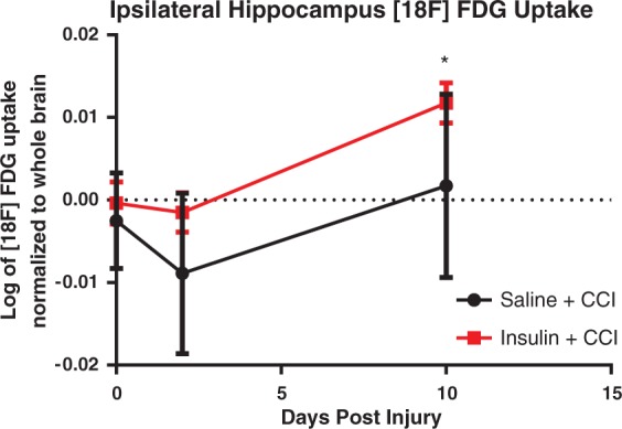
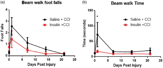
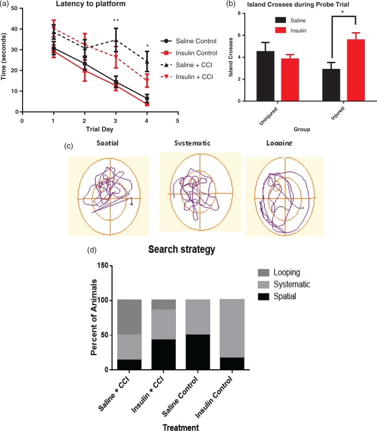
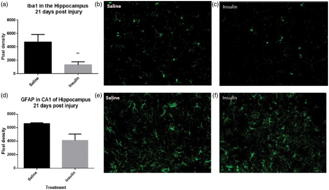
Traumatic brain injury (TBI) results in learning and memory dysfunction. Cognitive deficits result from cellular and metabolic dysfunction after injury, including decreased cerebral glucose uptake and inflammation. This study assessed the ability of intranasal insulin to increase cerebral glucose uptake after injury, reduce lesion volume, improve memory and learning function and reduce inflammation. Adult male rats received a controlled cortical impact (CCI) injury followed by intranasal insulin or saline treatment daily for 14 days. PET imaging of [18F]-FDG uptake was performed at baseline and at 48 h and 10 days post-injury and MRI on days three and nine post injury. Motor function was tested with the beam walking test. Memory function was assessed with Morris water maze. Intranasal insulin after CCI significantly improved several outcomes compared to saline. Insulin-treated animals performed better on beam walk and demonstrated significantly improved memory. A significant increase in [18F]-FDG uptake was observed in the hippocampus. Intranasal insulin also resulted in a significant decrease in hippocampus lesion volume and significantly less microglial immunolabeling in the hippocampus. These data show that intranasal insulin improves memory, increases cerebral glucose uptake and decreases neuroinflammation and hippocampal lesion volume, and may therefore be a viable therapy for TBI.

Keywords: Glucose uptake; intranasal insulin; microglia; positron emission tomography; traumatic brain injury.


Figures

Figure 1.
Figure 1.
Intranasal insulin delivery after CCI acts primarily in the CNS. The timeline displays times at which animals underwent surgery, behavior training, PET/CT imaging, and sacrifice.(a) The dose of intranasal insulin given was not sufficient to cause a significant change in blood glucose following CCI in intranasal insulin-treated animals (n = 4) compared to saline-treated injured controls (n = 4) as measured by tail vein blood draws at baseline, after injury/immediately prior to treatment and 3 h after treatment.(b) There was also no effect of intranasal insulin on body weight, measured through 21 days post-injury with 14 days of daily administration (n = 7/group injured n = 4/group uninjured; (c). CCI injury does not inhibit the pathway of intranasal administration to brain regions. (d) There was no significant difference between the ipsilateral and contralateral brain region and insulin was detected in the olfactory bulb, cerebellum, brain stem, hippocampus, and cortex 45 min after administration (n = 4). Data are represented as mean ± SEM.
Figure 2.
Figure 2.
Intranasal insulin decreased lesion volume in the hippocampus. Representative images at nine days post injury (a) from saline treated (left panel) and insulin treated (right panel. The colored ROIs indicate: red = uninjured control lesion area, green = injured lesion area, blue = edema. Total cortical lesion volume is significantly reduced over time, but there is no significant effect of treatment. (b, two-way ANOVA) There was no significant difference in cortex lesion volume (specific for edema, blue) between saline and insulin treated at days 3 or 9 post injury. (c, two-tailed t test) There was a significant decrease in hippocampus lesion volume in insulin-treated animals between days 3 and 9. (d, two-tailed t test *p < 0.05) There is no significant difference in hippocampal volume with insulin treatment over time. (e, two-tailed t test).
Figure 3.
Figure 3.
PET imaging of [18F]-FDG reveals significant differences in uptake patterns following intranasal insulin treatment compared to intranasal saline-treated animals after CCI (n = 5/group). PET scans of [18F]-FDG uptake were obtained with a CT for anatomical localization of brain regions. Uptake values from these regions were obtained by aligning a rat atlas to the scans. Regional analysis revealed that at 10 days post injury animals treated with intranasal insulin treatment following CCI had a significantly higher [18F]-FDG uptake in the ipsilateral hippocampus (a) than at baseline (p = 0.0329) and 2 days post injury (p = 0.0189). Data are represented as mean ± SEM. *=p < 0.05.
Figure 4.
Figure 4.
Intranasal insulin treatment after injury improves some aspects of motor function. After injury, both groups of animals (n = 7/group) show a non-significant increase in footfalls indicating a deficit in motor function that recovers over time (a). However, animals treated with intranasal insulin after injury were able to cross the beam significantly faster than saline-treated rats (b). Uninjured animals (n = 4/per group) show no significant difference in time (a) or foot falls (b). Data are presented as an average of time and footfalls across the four widths of board the animals were trained to cross. Data are represented as mean ± SEM. *=p < 0.05.
Figure 5.
Figure 5.
Intranasal insulin treatment improved memory function in Morris water maze tests. After CCI rats treated with intranasal saline performed significantly worse than uninjured controls at day three (p = 0.0078) and day 4 (p = 0.0247) of training (a) (n = 6/group uninjured, n = 10/group injured). However, during the probe trial, the injured animals treated with intranasal insulin (n = 7) crossed the island location significantly more times (p = 0.0330) in those treated with intranasal saline (n = 8) (b). There was no significant difference in island crosses between the uninjured treatment groups (n = 6/group). Search strategy utilized by rats during the probe trial was analyzed and categorized into one of three major groups (spatial, systematic, and looping) as previously described. Representative patterns are shown (c), and number of animals utilizing each strategy during the probe trial was quantified. Saline and insulin-treated uninjured animals (control) showed mostly spatial and systematic search strategies (d). A greater proportion of animals treated with saline after CCI showed a reduction in spatial search strategy and an increase in looping search methods. Likewise, fewer animals that received intranasal insulin for 14 days after injury displayed looping behavior than saline-treated rats, and a return of spatial search strategy (p < 0.0001). *p < 0.05 **p < 0.001. Points represent mean ± SEM.
Figure 6.
Figure 6.
Treatment with intranasal insulin after CCI reduces macrophage/microglia but does not significantly reduce astrocyte activation. Quantification of Iba1, a marker of microglia and macrophages, revealed a significant reduction (p = 0.0089) (a) in cells in CA1 of the ipsilateral hippocampus in animals treated with intranasal insulin (c) compared to intranasal saline (b) (n = 3 per group). Quantification of GFAP, a cell surface marker of astrocytes, did not show a significant difference (p = 0.0770) (d) between insulin-treated (n = 4) (f) and saline-treated animals (n = 3) (e). Points represent mean ± SEM. * = p < 0.05.

References

    1. Rassovsky Y, Levi Y, Agranov E, et al. Predicting long-term outcome following traumatic brain injury (TBI). J Clin Exp Neuropsychol 2015; 37: 354–366. - PubMed
    1. Giza CC, Hovda DA. The new neurometabolic cascade of concussion. Neurosurgery 2014 . October75 Suppl 4): S24–S33. - PMC - PubMed
    1. Fridman EA, Beattie BJ, Broft A, et al. Regional cerebral metabolic patterns demonstrate the role of anterior forebrain mesocircuit dysfunction in the severely injured brain. Proc Natl Acad Sci U S A 201429; 111: 6473–6478. - PMC - PubMed
    1. Li J, Gu L, Feng DF, et al. Exploring temporospatial changes in glucose metabolic disorder, learning, and memory dysfunction in a rat model of diffuse axonal injury. J Neurotrauma [Research Support, Non-U.S. Gov’t] 2012; 29: 2635–2646. - PMC - PubMed
    1. Vespa P, Bergsneider M, Hattori N, et al. Metabolic crisis without brain ischemia is common after traumatic brain injury: A combined microdialysis and positron emission tomography study. J Cereb Blood Flow Metab 2005; 25: 763–774. - PMC - PubMed

MeSH terms