. 2017 Jan 6:7:39612.
doi: 10.1038/srep39612.

Scaling of titanium implants entrains inflammation-induced osteolysis

Affiliations

Scaling of titanium implants entrains inflammation-induced osteolysis

Michal Eger et al. Sci Rep. .

Abstract

With millions of new dental and orthopedic implants inserted annually, periprosthetic osteolysis becomes a major concern. In dentistry, peri-implantitis management includes cleaning using ultrasonic scaling. We examined whether ultrasonic scaling releases titanium particles and induces inflammation and osteolysis. Titanium discs with machined, sandblasted/acid-etched and sandblasted surfaces were subjected to ultrasonic scaling and we physically and chemically characterized the released particles. These particles induced a severe inflammatory response in macrophages and stimulated osteoclastogenesis. The number of released particles and their chemical composition and nanotopography had a significant effect on the inflammatory response. Sandblasted surfaces released the highest number of particles with the greatest nanoroughness properties. Particles from sandblasted/acid-etched discs induced a milder inflammatory response than those from sandblasted discs but a stronger inflammatory response than those from machined discs. Titanium particles were then embedded in fibrin membranes placed on mouse calvariae for 5 weeks. Using micro-CT, we observed that particles from sandblasted discs induced more osteolysis than those from sandblasted/acid-etched discs. In summary, ultrasonic scaling of titanium implants releases particles in a surface type-dependent manner and may aggravate peri-implantitis. Future studies should assess whether surface roughening affects the extent of released wear particles and aseptic loosening of orthopedic implants.


Figures
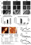
Figure 1
Figure 1. Topographic changes and titanium particles produced by US scaling.
Three different titanium surfaces, including M, SLA, and SB, were treated with US scaling on half of the disc area. (a) The border was examined using SEM at ×300 magnification. (b) The areas on both sides of the midline were examined at ×1500 magnification. In each panel, the left side represents the original disc surface, while the right side shows the disc after treatment. (c) Titanium particles released during this process were visualized using SEM to estimate their micro-roughness, (d) the number of particles and (e) the size distribution of the particles, which were evaluated using an automated cell counter. The size distribution is shown as a percentage of the total number of particles that originated from each surface type. (fh) Nanoroughness of particles was analyzed using atomic force microscopy (AFM). (f) Multiple line profiles were obtained for each particle type. (g) Slope, the fold-increase in linear distance and the average maximal distance between the highest and deepest points along 500 nm were calculated to determine particle nanoroughness. (h) Representative line profiles are shown for the different surfaces.
Figure 2
Figure 2. Titanium particles released from SLA titanium implants induce inflammatory responses in macrophages.
BMDMs were cultured for 24 hours with titanium particles that were released by ultrasonic scaling (Ti) and/or bacterial LPS (control). Saline was used as the control. IL1β, IL6 and TNFα expression levels were measured using RT-qPCR, normalized to β-actin and expressed as a fold-change relative to the control level. The data are shown as the mean ± SD of n = 5 for each condition. These data are from a representative experiment out of 5 *p < 0.05 versus control; **p < 0.05 versus LPS and the control.
Figure 3
Figure 3. Titanium particles stimulate osteoclastogenesis in vitro.
(a) TRAP staining (images and TRAP+area) of osteoclasts after 4 days of differentiation in the presence of LPS (0.01 μg/ml), titanium particles or diluent only (control). Bar = 30 μm. (b) The average number per well (N.Oc) and (c) the osteoclast area (mm2) in TRAP+cells after 4 days of differentiation. Data are shown as the mean ± SD. *p < 0.05 versus control; **p < 0.05 versus LPS and control.
Figure 4
Figure 4. Inflammatory response dependent on the number and origin of titanium particles.
BMDMs were cultured for 24 hours with: (a) Increasing numbers of titanium particles that were released by ultrasonic scaling of the SLA surface type. (b) 10% of the titanium particles that were released by ultrasonic scaling (Ti) of one disc from each implant surface: Machined (M), Sand-Blasted (SB), and SB/Acid-etched (SLA). (c) Same number of titanium particles (1293 particles/mm2) that were released by ultrasonic scaling (Ti) of each implant surface type: M, SB, SLA. IL1β, IL6 and TNFα expression levels were measured using RT-qPCR, normalized to β-actin and expressed as a fold-change relative to the control (no particles). The data are shown as the mean ± SD of n = 5 for each condition. These data are from a representative experiment out of 5. The regression analysis formula and goodness of fit (R2) are shown (a). *p < 0.05 versus M; **p < 0.05 versus SLA and M.
Figure 5
Figure 5. Schematic of exposed implant surfaces.
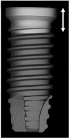
Micro-CT 3D rendering of a 3.75 mm diameter implant with a putative horizontal bone loss of 2.05 mm (arrows). The resulting exposed region (light gray) corresponds to a total implant surface of 28.27 mm2.
Figure 6
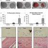
Figure 6. Titanium particles originating from US scaling of dental implants induce osteolysis in vivo.
Titanium particles that originated from US scaling of SB and SLA discs were inserted into fibrin membranes and implanted onto the calvaria of 6 mice per group. Animals were then sacrificed after 5 weeks. (a) Representative μCT images of the calvaria are shown. The region of interest (ROI) is represented as dark gray, and the resorption pits are represented as red. (b) Pit Resorption Volume (PRV, μm3) and (c) PRV are shown relative to bone tissue volume inside the ROI (PRV/TV, %). The data are expressed as the mean ± SD, n = 6. *p < 0.05 versus control (membrane with no particles); **p < 0.05 versus SLA and control. (d) Histological TRAP-stained sections demonstrating the increase in lining osteoclasts (blue arrows). (e) HE staining demonstrating the presence of blood vessels (*), inflammatory cells and fibrous tissue, especially in the SB group. Original magnification ×40.

Similar articles

Cited by

References

    1. Esposito M., Coulthard P., Thomsen P. & Worthington H. V. Interventions for replacing missing teeth: different types of dental implants. Cochrane Database of Systematic Reviews, CD003815, doi: 10.1002/14651858.CD003815.pub2 (2005). - DOI - PubMed
    1. Heitz-Mayfield L. J. & Mombelli A. The therapy of peri-implantitis: a systematic review. International Journal of Oral & Maxillofacial Implants 29 Suppl, 325–345, doi: 10.11607/jomi.2014suppl.g5.3 (2014). - DOI - PubMed
    1. Heitz-Mayfield L. J., Needleman I., Salvi G. E. & Pjetursson B. E. Consensus statements and clinical recommendations for prevention and management of biologic and technical implant complications. International Journal of Oral & Maxillofacial Implants 29 Suppl, 346–350, doi: 10.11607/jomi.2013.g5 (2014). - DOI - PubMed
    1. Mombelli A., Muller N. & Cionca N. The epidemiology of peri-implantitis. Clinical oral implants research 23 Suppl 6, 67–76, doi: 10.1111/j.1600-0501.2012.02541.x (2012). - DOI - PubMed
    1. Esposito M., Grusovin M. G. & Worthington H. V. Interventions for replacing missing teeth: treatment of peri-implantitis. Cochrane Database of Systematic Reviews 1, CD004970, doi: 10.1002/14651858.CD004970.pub5 (2012). - DOI - PMC - PubMed

Publication types

LinkOut - more resources