. 2017 Aug 18;8(6):875-890.
doi: 10.1080/21505594.2016.1278334. Epub 2017 Jan 6.

Molecular characterization of pneumococcal surface protein K, a potential pneumococcal vaccine antigen

Affiliations

Molecular characterization of pneumococcal surface protein K, a potential pneumococcal vaccine antigen

A-Yeung Jang et al. Virulence. .

Abstract

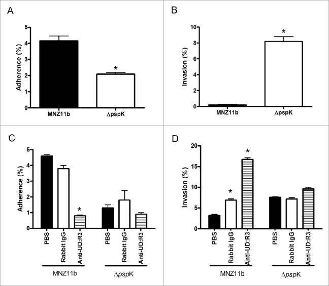
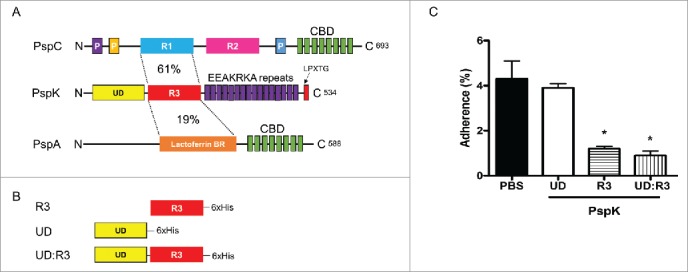
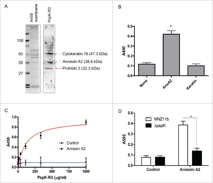
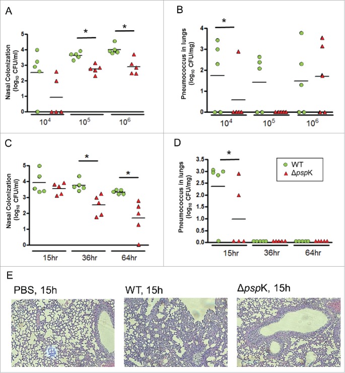
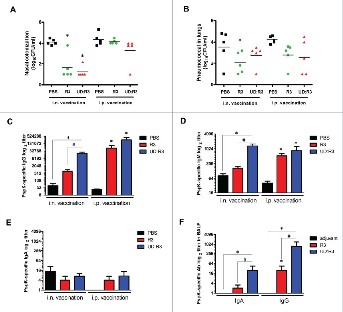
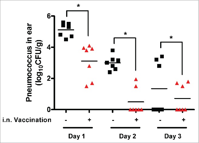
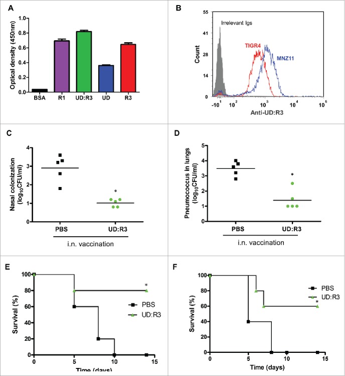
The pneumococcal capsule is indispensable for pathogenesis in systemic infections; however, many pneumococcal diseases, including conjunctivitis, otitis media, and some systemic infections in immunocompromised patients, are caused by nonencapsulated Streptococcus pneumoniae (NESp). Null capsule clade 1 (NCC1), found in group 2 NESp, expresses pneumococcal surface protein K (PspK) and is becoming prevalent among pneumococcal organisms owing to the widespread use of pneumococcal conjugate vaccines. Despite its clinical importance, the molecular mechanisms underlying the prevalence of NCC1 have not been fully elucidated. Here, we investigated the role of the R3 domain of PspK in the epithelial cell adherence of NCC1. We found that the R3 domain of PspK mediated NCC1 adherence via its direct interaction with the epithelial surface protein annexin A2. Additionally, neutralization with purified recombinant PspK-R3 or rabbit anti-UD:R3 IgG inhibited binding of NESp to lung epithelial cells in vitro. Immunization with the 'repeat' domain of PspK-R3 or PspK-UD:R3 effectively elicited mucosal and systemic immune responses against PspK-R3 and provided protection against nasopharyngeal, lung, and middle ear colonization of NESp in mice. Additionally, we found that rabbit anti-UD:R3 IgG bound to PspC-R1 of the encapsulated TIGR4 strain and that UD:R3 immunization provided protection against nasopharyngeal and lung colonization of TIGR4 and deaths by TIGR4 and D39 in mice. Further studies using 68 pneumococcal clinical isolates showed that 79% of clinical isolates showed cross-reactivity to rabbit anti-UD:R3 IgG. About 87% of serotypes in the 13-valent pneumococcal conjugate vaccine (PCV) and 68% of non-vaccine serotypes were positive for cross-reactivity with rabbit anti-UD:R3 IgG. Thus, the R3 domain of PspK may be an effective vaccine candidate for both NESp and encapsulated Sp.

Keywords: Streptococcus pneumoniae; annexin A2; pneumococcal surface protein K; vaccine.


Figures

Figure 1.
Figure 1.
PspK-mediated MNZ11b adherence to and invasion in human alveolar epithelial cells. ((A)& B) Adherence (A) and invasion (B) of WT MNZ11b or its isogenic ΔpspK mutant to A549 cells following infection at an MOI of 10 were assessed. ((C)& D) The effects of neutralization with rabbit anti-UD:R3 IgG on the adherence (C) and invasion (D) of WT and ΔpspK to A549 cells were assessed. Values are expressed as the total cell-associated CFU recovered compared with that of the original inoculum. Experiments were performed in triplicate and repeated at least twice. A representative experiment is shown. Data were analyzed by Student's t tests. *, p < 0.05 compared with MNZ11b in (A)and (B)and PBS in (C)and D.
Figure 2.
Figure 2.
Identification of the PspK domain mediating MNZ11b binding to A549 cells. (A) Domain organization of PspK and its homologs. The domains of PspK were compared with those of its closest homologs, PspC and PspA, using the NCBI BLAST program. Levels of amino acid identity between regions are indicated. (B) Alignment of the truncated form of PspK for purification. P: proline-rich region; R1, R2, and R3: ‘repeat’ domains; CBD: choline-binding domain; UD: undefined domain; BR: binding region; 6×His: 6 histidine tag. (C) Effects of pre-incubation with recombinant PspK domain fragments (UD, R3, or UD:R3) on MNZ11b binding to A549 cells. Values are expressed as the total cell-associated CFU recovered compared with that of the original inoculum. Experiments were performed in triplicate and repeated at least twice. A representative experiment is shown. Data were analyzed by Student's t tests. *, p < 0.05 compared with PBS.
Figure 3.
Figure 3.
Binding of PspK-R3 to annexin A2 on the A549 cell surface. (A) Far western blotting of cell membrane proteins from A549 cells with FLAGR3. Proteins were separated by SDS-PAGE on 10% gels (A549; left panel) and transferred to PVDF membranes. Proteins were then probed with FLAGR3 (10 µg/mL, right panel). Bound FLAGR3 was detected with anti-FLAG IgG. (B) Binding of FLAGR3 to annexin A2 or keratin was assessed by ELISA-based binding assays. (C) Binding curve of FLAGR3 to immobilized annexin A2. X-axis indicates amount of applied FLAGR3. (D) MNZ11b binding to annexin A2 was compared with its isogenic mutant ΔpspK binding to the wells immobilized with annexin A2 (10 µg per well). *, p < 0.05 compared with the None. None, the wells immobilized with casein blocking reagent.
Figure 4.
Figure 4.
Role of PspK expression in pneumococcal colonization and invasion. ((A)& B) Mice were inoculated with the indicated numbers of WT MNZ11b or ΔpspK cells intranasally (i.n.), and the numbers of colonized bacteria in the nasopharyngeal tract (A) and lungs (B) at 15 h postinfection (hpi) were counted. ((C)& D) Mice were i.n. inoculated with 105 CFU of WT or ΔpspK, and the numbers of colonized bacteria in the nasopharyngeal tract (C) and lungs (D) at 15, 36, and 64 h after inoculation were analyzed. The horizontal lines denote the median number of bacteria in each group of 5 mice. (E) Histopathology of representative lungs from mice infected with WT or ΔpspK (H&E staining). *, p < 0.05 compared with WT.
Figure 5.
Figure 5.
Protection against pneumococcal infection by immunization with PspK truncates. ((A)& B) Mice were immunized with 10 μg of UD:R3 or R3 proteins intranasally (i.n.) or intraperitoneally (i.p.), followed by i.n. challenge with 105 CFU MNZ11b. At 15 h postinfection (hpi), the numbers of bacteria in the nasopharynx (A) and lung (B) were counted from nasal washes or lung homogenates, respectively. (C–E) Serum levels of R3-specfic IgG (C), IgM (D), and IgA (E) were analyzed at 7 d after immunization. (F) R3-specific mucosal IgA and IgG in the BALF were analyzed. *, p < 0.05 compared with adjuvant; #, p < 0.05 compared with R3.
Figure 6.
Figure 6.
Protection against pneumococcal otitis media by immunizing PspK-R3. Mice were intranasally (i.n.) immunized twice at 10-day intervals with 50 μL PBS containing 10 μg PspK-R3 and 1 μg cholera toxin. Four days after the second immunization, R3-immunized mice and control mice inoculated with PBS were challenged with 2 × 10⁷ CFU of MNZ11b via the tympanic membrane. After 1, 2, and 3 d postinfection, the bullae of mice were dissected from the skull, middle ear washes were collected, and bacteria numbers in the middle ear cavity were counted by culturing serially diluted middle ear washes. *, p < 0.01.
Figure 7.
Figure 7.
Cross-protection against pneumococcal infection by immunization with UD:R3. (A) Rabbit anti-UD:R3 IgG diluted in PBS was incubated in 96-well plates containing 1ug/mL of immobilized purified R1, UD:R3, UD, or R3. The binding activity of rabbit anti-UD:R3 IgG to R1 was compared with reactivity to UD:R3, UD, and R3. (B) TIGR4 or NMZ11 was incubated with rabbit anti-UD:R3 IgG, followed by staining with FITC-conjugated goat anti-rabbit IgG, and FITC-positive pneumococci were analyzed by flow cytometry. ((C)& D) Mice were intranasally (i.n.) immunized with 10 μg of UD:R3 with 1 μg of cholera toxin, followed by challenge with 105 CFU of TIGR4. At 15 h postinfection (hpi), the numbers of bacteria in the nasopharynx (C) and lungs (D) were counted. ((E)& F) Mice were i.n. immunized with 10 μg of UD:R3 with 1 μg of cholera toxin, followed by challenge with 105 CFU of TIGR4 (E) or D39 (F). Survival of mice were monitored for 14 d. *, p < 0.01.

Comment in

Similar articles

Cited by

References

    1. Zhu F, Hu Y, Li J, Ye Q, Young MM Jr., Zhou X, Chen Z, Yan B, Liang JZ, Gruber WC, et al.. Immunogenicity and safety of 13-valent pneumococcal conjugate vaccine compared with 7-valent pneumococcal conjugate vaccine among healthy infants in China. Pediatr Infect Dis J 2016; :999-1010; PMID:27254028; http://dx.doi.org/10.1097/INF.0000000000001248 - DOI - PubMed
    1. Welte T. Pneumococcal conjugate vaccine-equally effective for everyone? Dtsch Arztebl Int 2016; 113(9):137-8. PMID:26987461 - PMC - PubMed
    1. van Werkhoven CH, Hollingsworth RC, Huijts SM, Bolkenbaas M, Webber C, Patterson S, Sanders EA, Bonten MJ. Pneumococcal conjugate vaccine herd effects on non-invasive pneumococcal pneumonia in elderly. Vaccine 2016; 34(28):3275-82; PMID:27171754; http://dx.doi.org/10.1016/j.vaccine.2016.05.002 - DOI - PubMed
    1. Valente C, Hinds J, Gould KA, Pinto FR, de Lencastre H, Sa-Leao R. Impact of the 13-valent pneumococcal conjugate vaccine on Streptococcus pneumoniae multiple serotype carriage. Vaccine 2016; 34(34):4072-8; PMID:27325351; http://dx.doi.org/10.1016/j.vaccine.2016.06.017 - DOI - PubMed
    1. Grant LR, Hammitt LL, O'Brien SE, Jacobs MR, Donaldson C, Weatherholtz RC, Reid R, Santosham M, O'Brien KL. Impact of the 13-valent pneumococcal conjugate vaccine on pneumococcal carriage among American Indians. Pediatr Infect Dis J 2016; 35(8):907-14; PMID:27171679; http://dx.doi.org/10.1097/INF.0000000000001207 - DOI - PMC - PubMed

MeSH terms