Review
. 2017 Jun;30(3):205-215.
doi: 10.1177/1971400916682752. Epub 2017 Jan 6.

Imaging of sialadenitis

Affiliations
Review

Imaging of sialadenitis

Ahmed Abdel Khalek Abdel Razek et al. Neuroradiol J. 2017 Jun.

Abstract

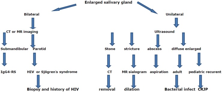
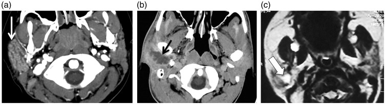
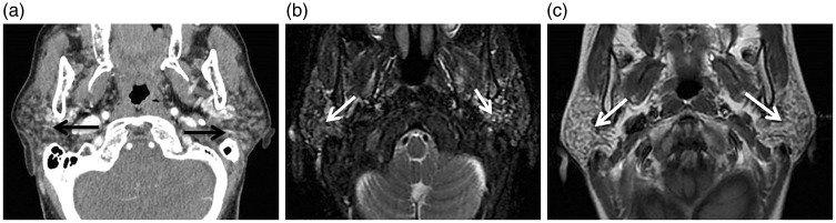
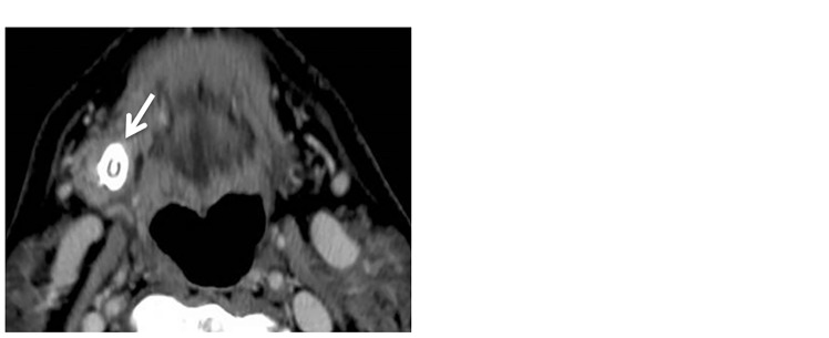
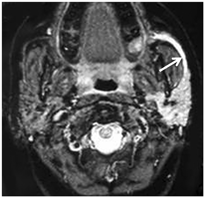
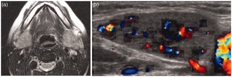
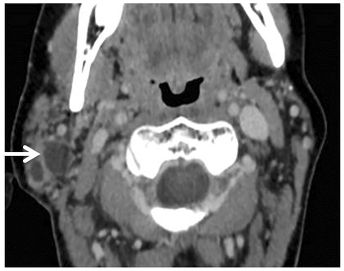
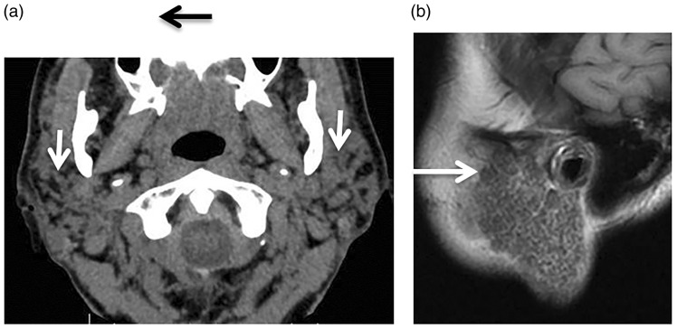
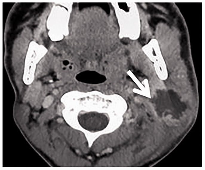
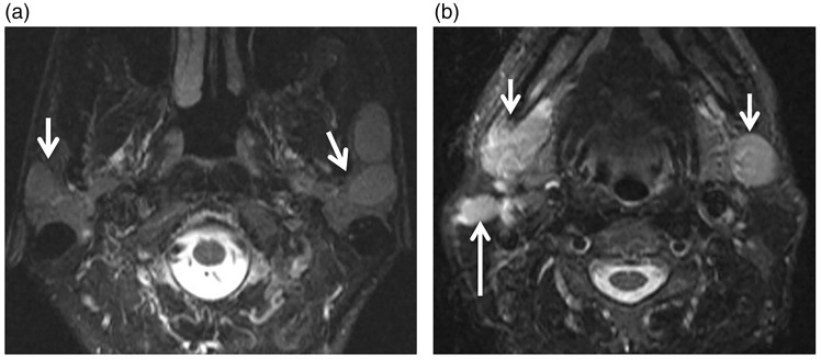
Sialadenitis is an inflammation or infection of the salivary glands that may affect the parotid, submandibular and small salivary glands. Imaging findings vary among unilateral or bilateral salivary gland enlargement, atrophy, abscess, ductal dilation, cysts, stones and calcification. Imaging can detect abscess in acute bacterial suppurative sialadenitis, ductal changes with cysts in chronic adult and juvenile recurrent parotitis. Imaging is sensitive for detection of salivary stones and stricture in obstructive sialadenitis. Immunoglobulin G4-sialadenitis appears as bilateral submandibular gland enlargement. Imaging is helpful in staging and surveillance of patients with Sjögren's syndrome. Correlation of imaging findings with clinical presentation can aid diagnosis of granulomatous sialadenitis. Post-treatment sialadenitis can occur after radiotherapy, radioactive iodine or surgery.

Keywords: Parotid; Sjögren’s; salivary; sialadenitis; stone; submandibular.


Figures

Figure 1.
Figure 1.
Flowchart algorithm showing the imaging approach for reaching a specific diagnosis of sialadenitis. CT: computed tomography; MR: magnetic resonance; IgG4-RS: immunoglobulin G4-related sialadenitis; HIV: human immunodeficiency virus; CRJP: chronic recurrent juvenile parotitis.
Figure 2.
Figure 2.
Acute sialadenitis. (a) Axial contrast computed tomography (CT) scan shows enlarged right parotid gland with intense enhancement (arrow). (b) Axial contrast CT scan of another patient shows a well-defined marginal enhanced abscess (arrow) in the right parotid gland. (c) Axial T2-weighted image shows hyperintense fluid with thick wall of abscess in the right parotid gland of a neonate (arrow).
Figure 3.
Figure 3.
Chronic adult sialadenitis. (a) Axial computed tomography (CT) shows granular appearance of both parotid glands (arrows). (b) Axial T2-weighted image shows multiple small cysts in both parotid glands (arrows). (c) Axial contrast T1-weighted image shows mild inhomogeneous pattern of enhancement of both parotid glands (arrows).
Figure 4.
Figure 4.
Chronic juvenile recurrent sialadenitis. Axial T2-weighted image shows an enlarged left parotid gland with mixed pattern of signal intensity (arrow).
Figure 5.
Figure 5.
Stone of submandibular duct. (a) Axial contrast computed tomography (CT) scan shows a stone (arrow) in the right submandibular gland region. (b) Axial T2-weighted image shows a signal void of a stone (arrow) in the right submandibular gland.
Figure 6.
Figure 6.
Obstructive sialadenitis. Axial gradient-recalled echo magnetic resonance (MR) image shows a dilated duct (long arrow) of the left parotid gland that exhibits high-signal intensity of the retained fluid.
Figure 7.
Figure 7.
Immunoglobulin G4-related sialadenitis (IgG4-RS). (a) Axial T2-weighted image shows both enlarged submandibular glands (arrow) that exhibit high signal intensity. (b) Color Duplex examination shows enlarged submandibular glands gland with increased color signal (arrow) and vascularity.
Figure 8.
Figure 8.
Human immunodeficiency virus (HIV) sialadenitis. Axial contrast computed tomography (CT) scan shows benign lymphoepithelial lesions (BLEBs) (arrow) of the right parotid gland in a patient with HIV.
Figure 9.
Figure 9.
Sjögren’s syndrome. (a) Axial contrast computed tomography (CT) scan shows granular appearance (arrow) of both parotid glands. (b) Sagittal T1-weighted image shows honey-comb appearance (arrow) of both parotid glands.
Figure 10.
Figure 10.
Tuberculous sialadenitis. Axial contrast computed tomography (CT) scan shows an enlarged right parotid gland with small marginally enhanced abscess (arrow).
Figure 11.
Figure 11.
Sarcoidosis. (a) Axial T2-weighted image shows well-defined hyperintense focal lesions (arrows) in both parotid glands more on the left side. (b) Axial T2-weighted image at another level shows enlarged both submandibular glands (short arrows) more on the right side with enlarged adjacent cervical lymph (long arrow).

References

    1. Mandel L. Salivary gland disorders. Med Clin North Am 2014; 98: 1407–1449. - PubMed
    1. Francis CL, Larsen C. Pediatric sialadenitis. Otolaryngol Clin North Am 2014; 47: 763–778. - PubMed
    1. Carlson ER. Diagnosis and management of salivary gland infections. Oral Maxillofac Surg Clin North Am 2009; 21: 293–312. - PubMed
    1. Thomas BL, Brown JE, McGurk M. Salivary gland disease. Front Oral Bio 2010; 14: 129–146. - PubMed
    1. Zenk J, Iro H, Klintworth N, et al. Diagnostic imaging in sialadenitis. Oral Maxillofacial Surg Clin North Am 2009; 21: 275–292. - PubMed

LinkOut - more resources