

Pseudoaneurysm formation due to rupture of intracranial aneurysms: Case series and literature review
- PMID: 28059632
- PMCID: PMC5433584
- DOI: 10.1177/1971400916684667
Pseudoaneurysm formation due to rupture of intracranial aneurysms: Case series and literature review
Abstract
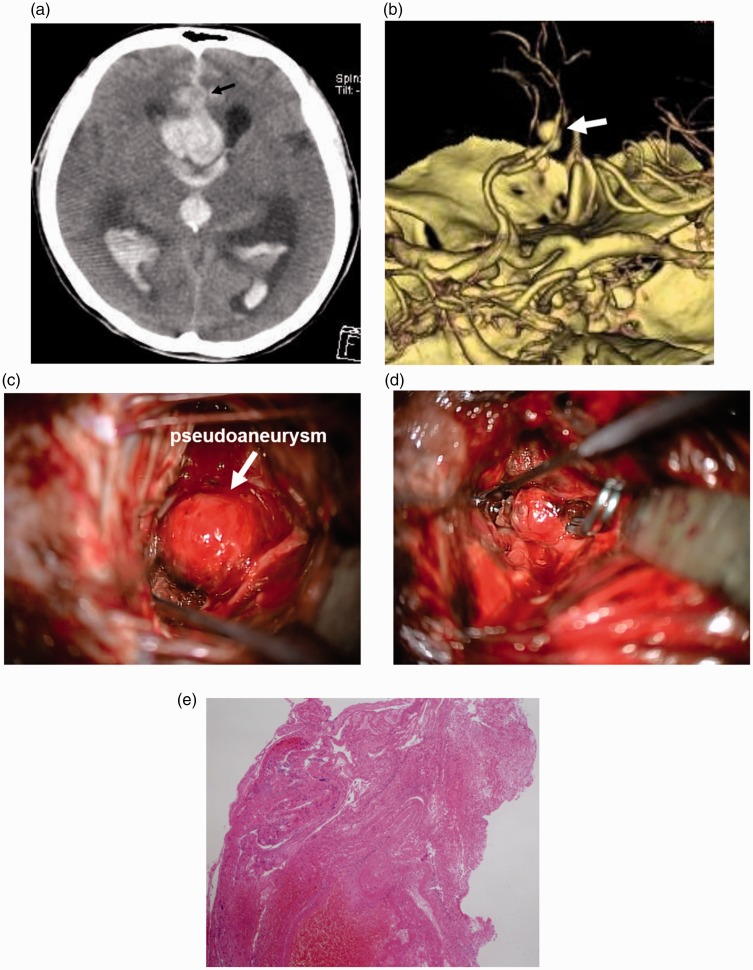
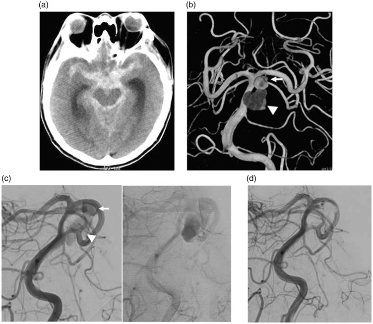
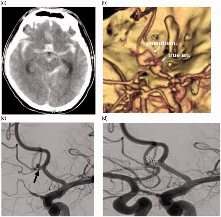
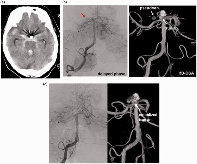
Background Intracranial pseudoaneurysm formation due to a ruptured non-traumatic aneurysm is extremely rare. We describe the radiological findings and management of pseudoaneurysms due to ruptured cerebral aneurysms in our case series and previously reported cases. Patients and methods Four additional and 20 reported patients presenting with subarachnoid hemorrhage (SAH) are included. Radiological findings and clinical features of these patients were reviewed. Results In our series, three-dimensional computed tomographic angiography (3D-CTA) and/or angiography showed an irregular- or snowman-shaped cavity extending from the parent artery. The radiological examination additionally revealed delayed filling and retention of contrast medium. These findings were the same as previously reported cases. One patient underwent direct clipping of the true aneurysm. For the other three patients with aneurysms at the basilar and anterior communicating arteries, the true portion of the aneurysm was embolized with platinum coils. During the procedures, care was taken not to insert the coils into the distal pseudoaneurysm portion to prevent rupture. The review of 24 cases revealed that the location of the aneurysms was most frequent in the anterior communicating artery (41.7%), and 86.7% of patients were in a severe stage of SAH (>Grade 3 in WFNS or Hunt & Kosnik grading) implying abundant SAH. Conclusions Pseudoaneurysm formation in SAH after non-traumatic aneurysm rupture is rare. However, in cases with an irregular-shaped aneurysm cavity, pseudoaneurysm formation should be taken into consideration.
Keywords: Pseudoaneurysm; cerebral aneurysm; intracerebral hematoma; subarachnoid hemorrhage.
Figures
References
-
- Teitelbaum GP, Dowd CF, Larsen DW, et al. Endovascular management of biopsy-related posterior inferior cerebellar artery pseudoaneurysm. Surg Neurol 1995; 43: 357–359. - PubMed
-
- Nomura M, Kida S, Uchiyama N, et al. Ruptured irregular shaped aneurysm: Pseudoaneurysm formation in a thrombus located at the rupture site. J Neurosurg 2000; 93: 998–1002. - PubMed
-
- Ide M, Kobayashi T, Tamano Y, et al. Pseudoaneurysm formation at the rupture site of a middle cerebral artery aneurysm. Neurol Med Chir (Tokyo) 2003; 43: 443–446. - PubMed
-
- Mori K, Kasuya C, Nakao Y, et al. Intracranial pseudoaneurysm due to rupture of a saccular aneurysm mimicking a large partially thrombosed aneurysm (“ghost aneurysm”): Radiological findings and therapeutic implications in two cases. Neurosurg Rev 2004; 27: 289–293. - PubMed
Publication types
MeSH terms
LinkOut - more resources
Full Text Sources
Other Literature Sources
Medical
