. 2017 Feb 17;355(6326):748-752.
doi: 10.1126/science.aai8792. Epub 2017 Jan 5.

Regeneration of fat cells from myofibroblasts during wound healing

Affiliations

Regeneration of fat cells from myofibroblasts during wound healing

Maksim V Plikus et al. Science. .

Abstract

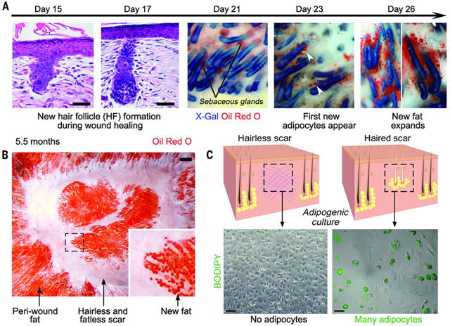
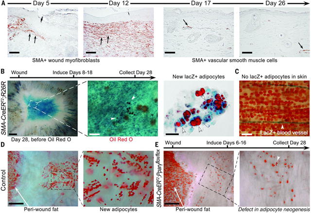
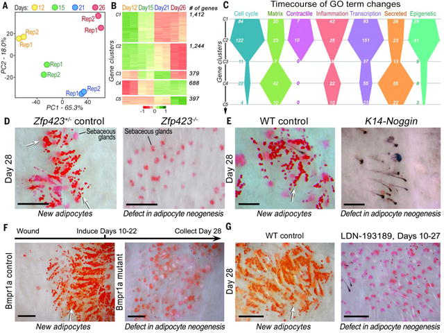
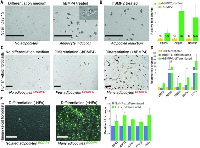
Although regeneration through the reprogramming of one cell lineage to another occurs in fish and amphibians, it has not been observed in mammals. We discovered in the mouse that during wound healing, adipocytes regenerate from myofibroblasts, a cell type thought to be differentiated and nonadipogenic. Myofibroblast reprogramming required neogenic hair follicles, which triggered bone morphogenetic protein (BMP) signaling and then activation of adipocyte transcription factors expressed during development. Overexpression of the BMP antagonist Noggin in hair follicles or deletion of the BMP receptor in myofibroblasts prevented adipocyte formation. Adipocytes formed from human keloid fibroblasts either when treated with BMP or when placed with human hair follicles in vitro. Thus, we identify the myofibroblast as a plastic cell type that may be manipulated to treat scars in humans.


Figures

Fig. 1
Fig. 1
New adipocytes only regenerate around new hair follicles during wound healing. (A) Histological sections (left two panels) and whole-mount images (right three panels) of skin stained to detect follicular epithelium (blue) and adipocytes (orange) in K14-Cre;R26R mice at indicated postwounding days. New adipocytes (arrowheads) increase in number and size over several days. (B) Skin viewed from the undersurface. New adipocytes form and persist exclusively around regenerated hair follicles, which arise in the center of the wound. (C) Cultured dermal cells isolated from wounds with regenerated hair follicles differentiated into BODIPY-positive (green) adipocytes, whereas cultured dermal cells from wounds lacking follicles formed no adipocytes. Scale bars in (A) and (C), 20 mm; in (B), 1 mm.
Fig. 2
Fig. 2
New adipocytes originate from wound myofibroblasts. (A) Smooth muscle actin (SMA)– positive myofibroblasts are present at the wound edge and then in the healing wound (arrows). By day 17, dermal wound cells express very little SMA, but vascular smooth muscle cells remain labeled (arrow). (B) Lineage tracing of myofibroblasts results in lacZ (blue)–expressing regenerated adipocytes (orange, white arrowheads). (C) Adipocytes in normal skin are not labeled. (D and E) Deletion of Pparg in myofibroblasts resulted in near-complete loss of new adipocytes, whereas normal cutaneous adipocytes at the wound edge remained intact. Scale bars in (A), 100 mm; in (B) (left), (D), and (E), 1 mm; in (B) (center), 200 mm; in (B) (right), 50 mm; in (C), 200 mm
Fig. 3
Fig. 3
Molecular profiling and functional studies of adipocyte regeneration reveal that ZFP423 and BMP signaling are necessary for adipocyte regeneration.(A) Principal component analysis of the myofibroblast transcriptome reveals distinct changes across four postwounding time points. (B) Differentially expressed genes (4120 total) from myofibroblasts at days 12 to 26 group into five distinct clusters (table S5). (C) Differentially expressed genes in several gene ontologies (GOs) undergo distinct temporal changes in myofibroblasts. (D) Deletion of Zfp423, (E) overexpression of the soluble BMP antagonist Noggin in K14-Noggin mice, (F) SMA-CreERT2–driven deletion of BMPR1A, and (G) treatment with the BMP antagonist LDN-193189 (2 mg/kg) during wound healing all resulted in near-complete loss of regenerated adipocytes in wounds, despite normal regeneration of hair follicles. WT, wild type. Scale bars in (D) to (G), 100 mm.
Fig. 4
Fig. 4
BMP drives reprogramming of mouse myofibroblasts and human keloid fibroblasts into adipocytes. (A and B) Treatment of cultured mouse dermal cells isolated from day-15 wounds with either human recombinant BMP4 or BMP2 induced their reprogramming into adipocytes and the activation of adipocyte-specific genes. Day-15 dermal cells cultured in pro-adipogenic differentiation media without BMP remained nonadipogenic. (C and D) Treatment of cultured human keloid scar cells with human recombinant BMP4 and adipocyte differentiation media induced their reprogramming into adipocytes with activation of adipocyte-specific genes. (E) In a coculture system, human scalp hair follicles induced adipogenic conversion of human keloid scar cells with (F) a concomitant increase in adipocyte genes. Scale bars in (A) and (B), 400 mm; in (C) and (E), 200 mm. Values in the graphs are means ± SEM. *P < 0.028; **P < 0.0002.

Comment in

References

    1. Gay D, et al. Fgf9 from dermal gammadelta T cells induces hair follicle neogenesis after wounding. Nature medicine. 2013;19:916–923. - PMC - PubMed
    1. Ito M, et al. Wnt-dependent de novo hair follicle regeneration in adult mouse skin after wounding. Nature. 2007;447:316–320. - PubMed
    1. Seifert AW, et al. Skin shedding and tissue regeneration in African spiny mice (Acomys) Nature. 2012;489:561–565. - PMC - PubMed
    1. Wang X, et al. Principles and mechanisms of regeneration in the mouse model for wound-induced hair follicle neogenesis. Regeneration (Oxf) 2015;2:169–181. - PMC - PubMed
    1. Breedis C. Regeneration of hair follicles and sebaceous glands from the epithelium of scars in the rabbit. Cancer Res. 1954;14:575–579. - PubMed

Publication types

MeSH terms