. 2017 Jan 6;355(6320):89-92.
doi: 10.1126/science.aah5163.

Cryo-EM structures and atomic model of the HIV-1 strand transfer complex intasome

Affiliations

Cryo-EM structures and atomic model of the HIV-1 strand transfer complex intasome

Dario Oliveira Passos et al. Science. .

Abstract

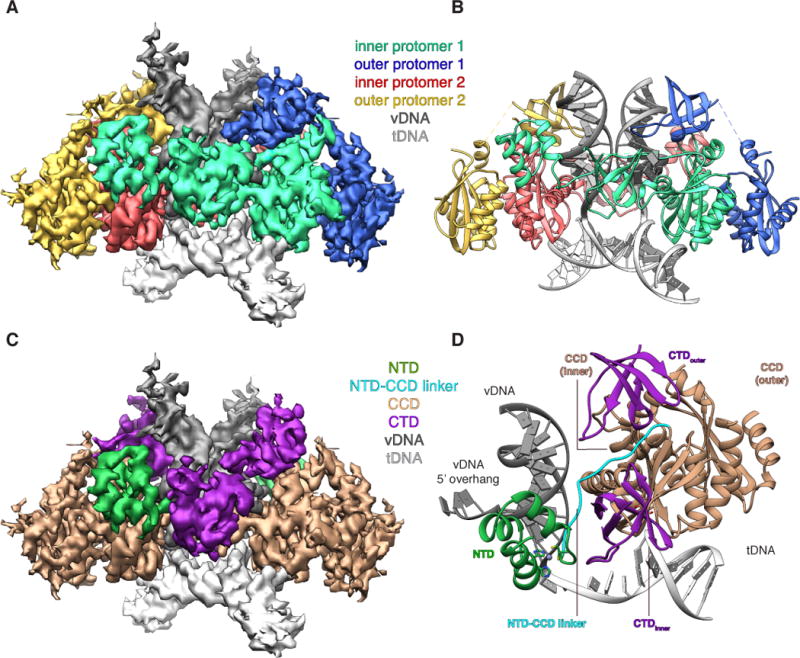
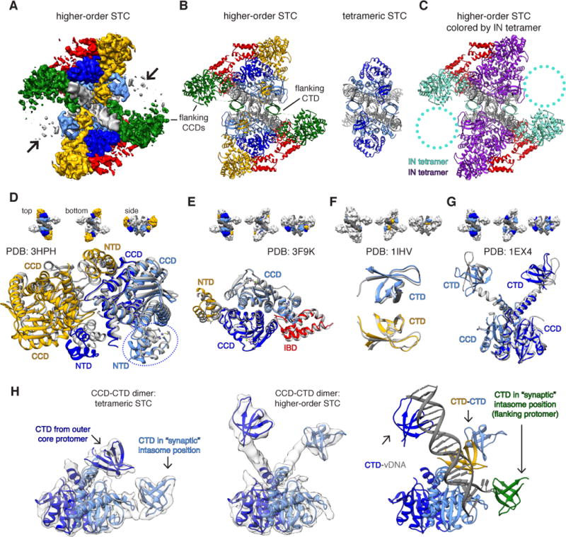
Like all retroviruses, HIV-1 irreversibly inserts a viral DNA (vDNA) copy of its RNA genome into host target DNA (tDNA). The intasome, a higher-order nucleoprotein complex composed of viral integrase (IN) and the ends of linear vDNA, mediates integration. Productive integration into host chromatin results in the formation of the strand transfer complex (STC) containing catalytically joined vDNA and tDNA. HIV-1 intasomes have been refractory to high-resolution structural studies. We used a soluble IN fusion protein to facilitate structural studies, through which we present a high-resolution cryo-electron microscopy (cryo-EM) structure of the core tetrameric HIV-1 STC and a higher-order form that adopts carboxyl-terminal domain rearrangements. The distinct STC structures highlight how HIV-1 can use the common retroviral intasome core architecture to accommodate different IN domain modules for assembly.


Figures

Fig. 1
Fig. 1. HIV-1 STC intasome structure
(A). CryoEM reconstruction of the STC, segmented by IN protomer (red, green, yellow, blue) and product DNA component (dark and light grey). (B) Atomic model derived from the cryoEM density, colored as in A. (C) segmented cryoEM density and (D) asymmetric subunit of the atomic model, colored by IN domain; NTD (green), CCD (beige), NTD-CCD linker (cyan), CTD (purple).
Fig. 2
Fig. 2. Network of interactions within the HIV-1 STC intasome
(A–C) Protein color scheme as in Fig. 1C–D. (A) Map of IN residues predicted to be involved in electrostatic protein-DNA interactions within the STC intasome structure. All three domains and the NTD-CCD linker participate in interactions with vDNA (pink), while tDNA interactions in blue are mostly restricted to the CCD. (B) Predicted inter-domain H-bond interactions within the STC. Residues designated below the domain schematic refer to the interacting domain and are colored accordingly. (C) Close-ups of selected regions involved in DNA interactions. For comparing HIV-1 R231 with PFV R329, the two structures were aligned to tDNA.
Fig. 3
Fig. 3. HIV-1 STC intasomes form higher order oligomers through distinct mechanisms of assembly
(A) CryoEM density map of IBD-bound STCs (STCIBD). Densities are segmented either by IN protomer (inner core: light blue, outer core: dark blue) or IN dimer (yellow and green), while IBD is in red. (B) Higher-order STC model assembled by rigid-body docking individual domain components, colored as in A. The higher-order STC (left) is shown side-by-side with the tetrameric STC from Fig. 1 (right). (C) Model as in B, colored by IN tetramer (28). Circled regions contain poorly resolved density that may harbor additional IN dimers. (DG) Structural comparison of higher-order STC assembled through rigid-body docking of individual domains with prior multi-domain IN structures. The structural components of higher-order STCs are colored as in panels AB, while the PDB structures used for comparison are gray. Comparisons include: (D) MVV INNTD-CCD tetramer (PDB: 3HPH, IBD has been omitted for clarity; the circled NTD arises from an IN protomer on the opposite side of vDNA), (E) HIV-2 INNTD-CCD dimer bound to IBD (PDB: 3F9K), (F) HIV-1 CTD dimer (PDB: 1IHV), and (G) HIV-1 INCCD-CTD dimer (PDB: 1EX4). (H) Conformational rearrangement within the core CCD-CTD dimer between (left) the tetrameric STC and (center) a higher-order STC, both overlaid on respective filtered experimental EM density. At right, the rearranged higher-order dimer is displayed in the context of additional CTDs and vDNA within the asymmetric unit. The “synaptic” position is required to form the conserved intasome core interface present in all retroviral intasomes.

References

    1. Craigie R, Bushman FD. HIV DNA integration. Cold Spring Harb Perspect Med. 2012;2:a006890–a006890. - PMC - PubMed
    1. Lesbats P, Engelman AN, Cherepanov P. Retroviral DNA Integration. Chem Rev. 2016 acs.chemrev.6b00125. - PMC - PubMed
    1. Blanco JL, Whitlock G, Milinkovic A, Moyle G. HIV integrase inhibitors: a new era in the treatment of HIV. Expert Opinion on Pharmacotherapy. 2015;16:1313–1324. - PubMed
    1. Dyda F, et al. Crystal structure of the catalytic domain of HIV-1 integrase: similarity to other polynucleotidyl transferases. Science. 1994;266:1981–1986. - PubMed
    1. Eijkelenboom AP, et al. The DNA-binding domain of HIV-1 integrase has an SH3-like fold. Nat Struct Biol. 1995;2:807–810. - PubMed

Publication types