. 2017 Jan 6;355(6320):93-95.
doi: 10.1126/science.aah7002.

A supramolecular assembly mediates lentiviral DNA integration

Affiliations

A supramolecular assembly mediates lentiviral DNA integration

Allison Ballandras-Colas et al. Science. .

Abstract

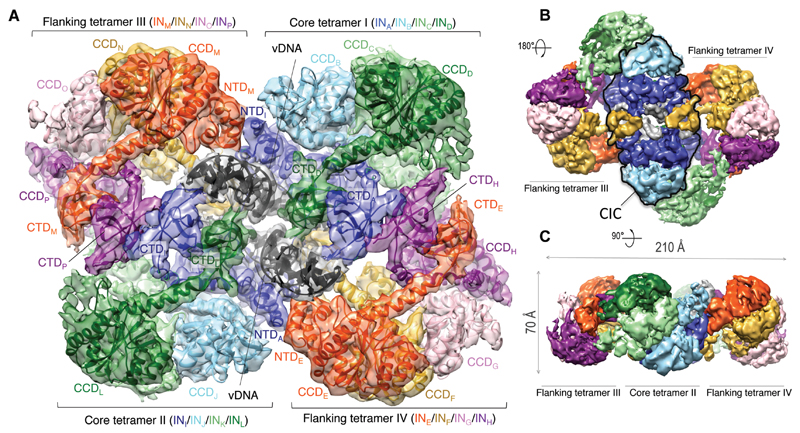
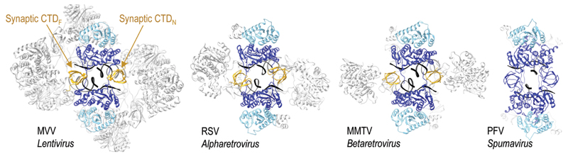
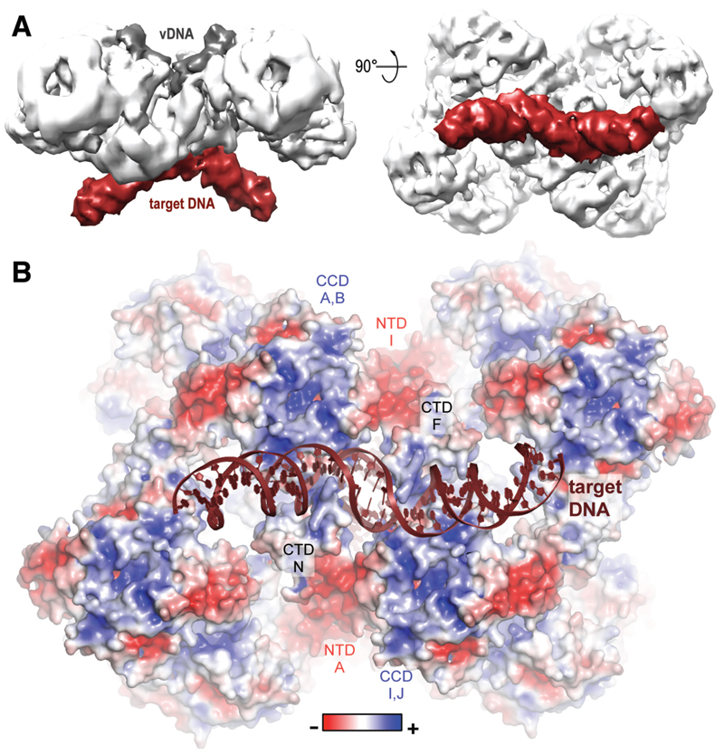
Retroviral integrase (IN) functions within the intasome nucleoprotein complex to catalyze insertion of viral DNA into cellular chromatin. Using cryo-electron microscopy, we now visualize the functional maedi-visna lentivirus intasome at 4.9 angstrom resolution. The intasome comprises a homo-hexadecamer of IN with a tetramer-of-tetramers architecture featuring eight structurally distinct types of IN protomers supporting two catalytically competent subunits. The conserved intasomal core, previously observed in simpler retroviral systems, is formed between two IN tetramers, with a pair of C-terminal domains from flanking tetramers completing the synaptic interface. Our results explain how HIV-1 IN, which self-associates into higher-order multimers, can form a functional intasome, reconcile the bulk of early HIV-1 IN biochemical and structural data, and provide a lentiviral platform for design of HIV-1 IN inhibitors.


Figures

Fig. 1
Fig. 1. Cryo-EM reconstruction of the MVV intasome.
A. Fitted intasome model color-coded to highlight IN subunits including 12 NTDs, 16 CCDs, and 14 CTDs. Molecules of vDNA in dark grey are surrounded by core tetramers I and II (colored in green, light green, sky blue, and blue), and flanking tetramers III and IV (red, yellow, pink, and purple). B-C. Views of the map in two alternative orientations. The CIC structure is highlighted with a black outline in panel B.
Fig. 2
Fig. 2. CIC structure in MVV and previously characterized synaptic complexes.
The CIC in each structure is shown in color with the remainder in grey; yellow CTDs indicate domains donated by flanking IN subunits.
Fig. 3
Fig. 3. Target DNA binding and surface electrostatic potential distribution.
A. Cryo-EM reconstruction of the MVV strand transfer complex at 8.6 Å resolution viewed in two orientations. Protein, vDNA, and target DNA are shown in white, dark grey and bordeaux, respectively. B. Pseudo-atomic model of the STC with the protein portion of the structure in space-fill mode and colored by charge.

References

    1. Lesbats P, Engelman AN, Cherepanov P. Retroviral DNA Integration. Chem Rev. 2016;116:12730–12757. - PMC - PubMed
    1. Hare S, Gupta SS, Valkov E, Engelman A, Cherepanov P. Retroviral intasome assembly and inhibition of DNA strand transfer. Nature. 2010;464:232–236. - PMC - PubMed
    1. Maertens GN, Hare S, Cherepanov P. The mechanism of retroviral integration from X-ray structures of its key intermediates. Nature. 2010;468:326–329. - PMC - PubMed
    1. Yin Z, Shi K, Banerjee S, Pandey KK, Bera S, Grandgenett DP, Aihara H. Crystal structure of the Rous sarcoma virus intasome. Nature. 2016;530:362–366. - PMC - PubMed
    1. Ballandras-Colas A, Brown M, Cook NJ, Dewdney TG, Demeler B, Cherepanov P, Lyumkis D, Engelman AN. Cryo-EM reveals a novel octameric integrase structure for betaretroviral intasome function. Nature. 2016;530:358–361. - PMC - PubMed

Publication types

Substances