Review
. 2017;35(1):87-103.
doi: 10.3233/RNN-160690.

Recent progress in translational research on neurovascular and neurodegenerative disorders

Collaborators, Affiliations
Free PMC article
Review

Recent progress in translational research on neurovascular and neurodegenerative disorders

Hans-Ulrich Demuth et al. Restor Neurol Neurosci. 2017.
Free PMC article

Abstract

The already established and widely used intravenous application of recombinant tissue plasminogen activator as a re-opening strategy for acute vessel occlusion in ischemic stroke was recently added by mechanical thrombectomy, representing a fundamental progress in evidence-based medicine to improve the patient's outcome. This has been paralleled by a swift increase in our understanding of pathomechanisms underlying many neurovascular diseases and most prevalent forms of dementia. Taken together, these current advances offer the potential to overcome almost two decades of marginally successful translational research on stroke and dementia, thereby spurring the entire field of translational neuroscience. Moreover, they may also pave the way for the renaissance of classical neuroprotective paradigms.This review reports and summarizes some of the most interesting and promising recent achievements in neurovascular and dementia research. It highlights sessions from the 9th International Symposium on Neuroprotection and Neurorepair that have been discussed from April 19th to 22nd in Leipzig, Germany. To acknowledge the emerging culture of interdisciplinary collaboration and research, special emphasis is given on translational stories ranging from fundamental research on neurode- and -regeneration to late stage translational or early stage clinical investigations.

Keywords: Alzheimer’s disease; brain; cerebral ischemia; cerebral small vessel disease; dementia; experimental therapy; hemorrhage; in vivo imaging; neuroimmunology; neuroprotection; neurorepair; sex differences; stroke; translational research; vascular cognitive impairment.


Figures

Fig.1
Fig.1
Increase of infarct core volume due to ICP-caused collateral supply decline. (A) Collateral perfusion helps to limit the extent of the ischemic core after stroke if ICP remains low to moderate. (B) Steeply increasing ICP can dramatically reduce collateral blood flow after ischemic stroke, since the cerebral perfusion pressure equals to the mean arterial pressure minus ICP as suggested by the Monro–Kellie doctrine. Challenging conventional knowledge suggesting that edema is the major cause of ICP increase, Murtha and colleagues could show that even minor ischemic lesions causing mild edema might lead to significant ICP rises. This may represent an important pathophysiological mechanism contributing to significant cerebral damage even after initial minor strokes with larger penumbras with good collateralization. A potential therapeutic strategy might be the application of mild hypothermia, which has been demonstrated to prevent the ICP rise after even minor strokes. Angiograms were taken from Shuaib et al., 2011.
Fig.2
Fig.2
Comparison between rodent, ovine and human brain. Although significantly smaller than the human brain, the ovine brain is also gyrencephalic and exhibits a grey-to-white-matter ration more similar to humans as compared to the rodent brains. Sheep are therefore considered an interesting model species for translational neuroscience.
Fig.3
Fig.3
Changes in neural network connectivity after stroke. (A) Schematic representation of a neural network in human brain before (left), with nodes and edges in blue, and after stroke (right), illustrating global disconnection (red dotted edges) and reorganization (green edges) as a result of a focal stroke lesion (black area). (B) Diffusion tensor imaging-based fiber tractography maps of control (left) and 10-weeks post-stroke rat brain (right), showing altered fiber pathway patterns in perilesional white matter chronically after stroke. Stroke was induced by 90-min intraluminal occlusion of the right middle cerebral artery in adult male Sprague Dawley rats. High angular (120 directions) and spatial resolution (0.2 mm isotropic voxels) diffusion-weighted MRI data were acquired post mortem on a 9.4 T animal MRI scanner (total scan time: 26 h). MrTrix3® software (http://www.mrtrix.org/) was used for diffusion-based tractography. Courtesy of Michel Sinke and Willem Otte.
Fig.4
Fig.4
Transcranial direct current stimulation (tDCS) induces neurogenesis and accelerates functional recovery after stroke in the rodent brain. (A) Neuroblasts in the subventricular zone (SVZ) were identified by their expression of doublecortin (DCX) under control conditions (sham, left) and after multi-session tDCS of cathodal (middle) or anodal (right) polarity. (B) Following multi-session tDCS of cathodal or anodal polarity, the area of DCX-positive neuroblasts in the SVZ ipsilateral to tDCS was wider than under control conditions in the healthy mouse brain (sham). (C) Rats subjected to focal cerebral ischemia by middle cerebral artery occlusion displayed a wider DCX+area of the SVZ when treated with tDCS of cathodal or anodal polarity for 10 consecutive days after stroke. (D) Motor recovery assessed by the Catwalk test revealed that multi-session cathodal tDCS led to a faster recovery of limb strength (‘print area’) in stroke rats compared to sham treatment. (E) Both cathodal and anodal tDCS facilitated recovery of gait, i.e., led to less limping (‘stand’), compared to control (*p < 0.05). Figure elements were modified from Pikhovych et al., 2016 and Braun et al., 2016 with permissions.

References

    1. Antonic A., Dottori M., Leung J., Sidon K., Batchelor P.E., Wilson W., Macleod M.R., & Howells D.W. (2014). Hypothermia protects human neurons. International Journal of Stroke, 9(5), 544–552. - PMC - PubMed
    1. Banerjee A., Wang J., Bodhankar S., Vandenbark A.A., Murphy S.J., & Offner H. (2013). Phenotypic changes in immune cell subsets reflect increased infarct volume in male vs. female mice. Translational Stroke Research, 4(5), 554–563. - PMC - PubMed
    1. Bauer A.Q., Kraft A.W., Wright P.W., Snyder A.Z., Lee J.M., & Culver J.P. (2014). Optical imaging of disrupted functional connectivity following ischemic stroke in mice. Neuroimage, 99, 388–401. - PMC - PubMed
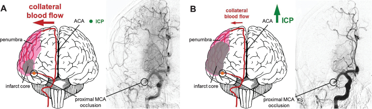
    1. Beard D.J., Murtha L.A., McLeod D.D., & Spratt N.J. (2016b). Intracranial pressure and collateral blood flow. Stroke, 47(6), 1695–1700. - PubMed
    1. Beard D.J., Logan C.L., McLeod D.D., Hood R.J., Pepperall D., Murtha L.A., & Spratt N.J. (2016a). Ischemic penumbra as a trigger for intracranial pressure rise - A potential cause for collateral failure and infarct progression? Journal of Cerebral Blood Flow and Metabolism, 36(5), 917–927. - PMC - PubMed

MeSH terms