

Patients With Dry Eye Disease and Low Subbasal Nerve Density Are at High Risk for Accelerated Corneal Endothelial Cell Loss
- PMID: 28060067
- PMCID: PMC5298224
- DOI: 10.1097/ICO.0000000000001057
Patients With Dry Eye Disease and Low Subbasal Nerve Density Are at High Risk for Accelerated Corneal Endothelial Cell Loss
Abstract
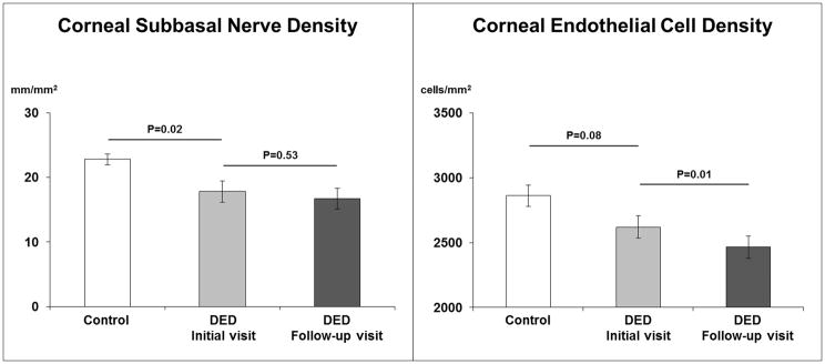
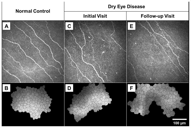
Purpose: To evaluate changes in corneal endothelial cell density over time in patients with dry eye disease (DED) and to correlate endothelial cell loss with corneal subbasal nerve density.
Methods: This retrospective study included 40 eyes of 20 patients with DED. Laser in vivo confocal microscopy had been performed in the central cornea of both eyes at an initial visit and repeated after a mean follow-up of 33.2 ± 10.2 months. The densities of corneal endothelial cells and subbasal nerves were measured in both visits and compared with 13 eyes of 13 normal age-matched controls.
Results: At the initial visit, the DED group had lower densities of corneal endothelial cells (2620 ± 386 cells/mm) and subbasal nerves (17.8 ± 7.5 mm/mm) compared with the control group (2861 ± 292 cells/mm and 22.8 ± 3.0 mm/mm, with P = 0.08 and P = 0.01, respectively). At the end of follow-up, although there was no significant change in subbasal nerve density (16.7 ± 7.2 mm/mm, P = 0.43), the mean corneal endothelial cell density significantly decreased to 2465 ± 391 cells/mm (P = 0.01), with a mean corneal endothelial cell loss of 2.1 ± 3.6% per year. The endothelial cell loss showed a statistically significant negative correlation with the initial subbasal nerve density (Rs = -0.55, P = 0.02).
Conclusions: Patients with DED have an accelerated corneal endothelial cell loss compared with that reported in the literature for normal aging. Those with lower subbasal nerve density, in particular, are at a higher risk for endothelial cell loss over time.
Figures
References
-
- The epidemiology of dry eye disease: report of the Epidemiology Subcommittee of the International Dry Eye WorkShop. Ocul Surf. 2007;5:93–107. - PubMed
-
- Lin PY, Tsai SY, Cheng CY, et al. Prevalence of dry eye among an elderly Chinese population in Taiwan: the Shihpai Eye Study. Ophthalmology. 2003;110:1096–1101. - PubMed
-
- McCarty CA, Bansal AK, Livingston PM, et al. The epidemiology of dry eye in Melbourne, Australia. Ophthalmology. 1998;105:1114–1119. - PubMed
-
- Villani E, Mantelli F, Nucci P. In-vivo confocal microscopy of the ocular surface: ocular allergy and dry eye. Curr Opin Allergy Clin Immunol. 2013;13:569–576. - PubMed
Publication types
MeSH terms
Grants and funding
LinkOut - more resources
Full Text Sources
Other Literature Sources
