Review
. 2017 Aug 23;117(16):10726-10759.
doi: 10.1021/acs.chemrev.6b00582. Epub 2017 Jan 6.

Watching Proteins Wiggle: Mapping Structures with Two-Dimensional Infrared Spectroscopy

Affiliations
Review

Watching Proteins Wiggle: Mapping Structures with Two-Dimensional Infrared Spectroscopy

Ayanjeet Ghosh et al. Chem Rev. .

Abstract

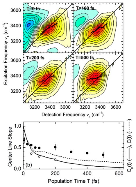
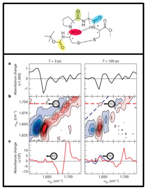
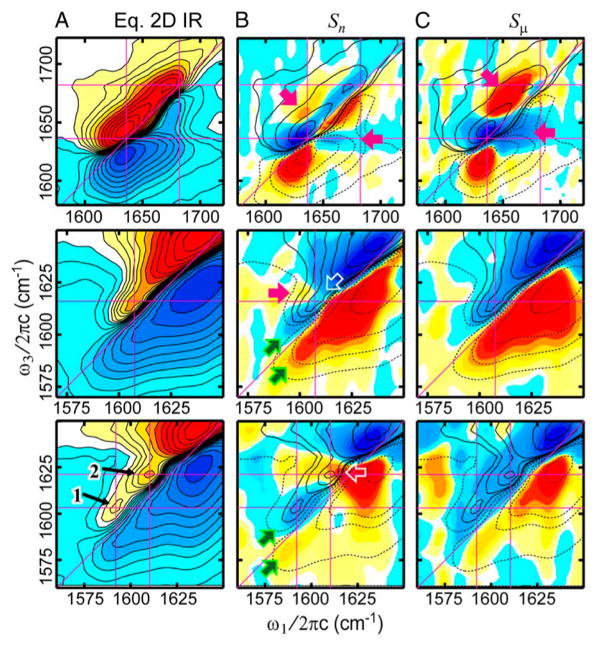
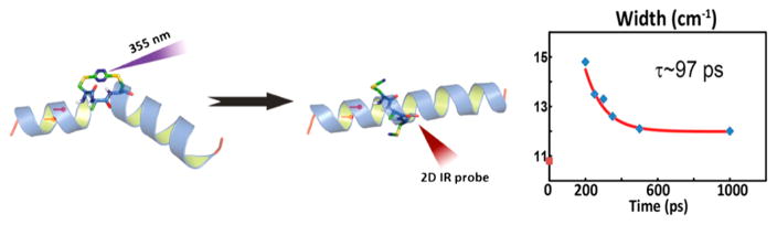
Proteins exhibit structural fluctuations over decades of time scales. From the picosecond side chain motions to aggregates that form over the course of minutes, characterizing protein structure over these vast lengths of time is important to understanding their function. In the past 15 years, two-dimensional infrared spectroscopy (2D IR) has been established as a versatile tool that can uniquely probe proteins structures on many time scales. In this review, we present some of the basic principles behind 2D IR and show how they have, and can, impact the field of protein biophysics. We highlight experiments in which 2D IR spectroscopy has provided structural and dynamical data that would be difficult to obtain with more standard structural biology techniques. We also highlight technological developments in 2D IR that continue to expand the scope of scientific problems that can be accessed in the biomedical sciences.


Conflict of interest statement

The authors declare the following competing financial interest(s): M.T.Z. is a co-owner of PhaseTech Spectroscopy, which manufactures mid-IR pulse shapers and 2D IR spectrometers.

Figures

Figure 1
Figure 1
Typical features of a 2D IR spectrum. (a) Diagonal peaks and (b) cross-peaks. The spectra shown were simulated from the third-order response functions described in ref .
Figure 2
Figure 2
(a) Simulated IR absorption spectra of 210–510 β-sheets. (b) The inverse participation ratios of each of the normal modes. (c) The Fα (M) spectrum, which is approximately a measure of the number of strands participating in a given normal mode. Reproduced with permissionfrom ref . Copyright 2004 American Chemical Society.
Figure 3
Figure 3
Effects of correlated line broadening on 2D line shapes for fully correlated (ρpq = +1) (a–c) and fully anticorrelated (ρpq = −1) (d–f) cases for rephasing (a, d), nonrephasing (b, e), and 2D correlation spectra (c, f). The simulations are done for the RDC six-level system assuming that the dynamics of system bath interactions are completely separable into fast (Γaa = Γss = Γas = 2 cm−1) and slow (σaa = σss = σas = 10 cm−1) components. Note that in the case of ρpq = +1 cross-peaks are tilted parallel to the diagonal in the rephasing spectrum (a), and the amplitudes of the cross-peaks are suppressed relative to diagonal peaks in the nonrephasing spectrum (b). For ρpq = −1, the cross-peaks are tilted perpendicular to the diagonal, and their amplitudes are enhanced relative to the diagonal peaks in the nonrephasing spectrum (e), whereas the cross-peak amplitudes are suppressed in the rephasing spectrum (d). The difference in the cross-peak intensities in the correlation spectra (c–f) is due to the imbalance in the number of corresponding pathways in the generation of the nonrephasing signal. The contours are plotted at 8% intervals. Reproduced with permission from ref . Copyright 2003 American Chemical Society.
Figure 4
Figure 4
Structure of the macrocyclic peptide with numbering scheme for isotope-labeled pairs (A). 2D IR data for the unlabeled macrocycle (B) and for the doubly labeled macrocycle (C–H). This figure demonstrates the shift observed upon isotope labeling the amide backbone. Reproduced with permission from ref . Copyright 2012 American Chemical Society.
Figure 5
Figure 5
Relaxation of the frequency autocorrelation function (FFCF) is reflected in the waiting time (T) evolution of 2D IR spectra: the spectra are more elongated at early T and become more symmetric at longer T as the FFCF decays. The correlation relaxation can be quantitatively measured through slope of the 2D contours as a function of T. (a) The nodal line slope and (b) the center line slope.
Figure 6
Figure 6
Calculated infrared (top) and 2D IR spectra (bottom) for an idealized extended β-sheet composed of a 3 × 3 lattice of the unit cells. Calculations are for (a) periodic boundary conditions and (b) open boundary conditions. The 2D IR calculations assumed a crossed polarization (XXYY) condition. Lorentzian line shapes are used with a width of (a) γ = 4 cm−1 and (b) γ = 2.5 cm−1. Positive features are shown in red, and negative features are shown in blue. Equally spaced contours are plotted between −10% and 10% of the a-diagonal peak maximum. Reproduced from ref . with permission. Copyright 2004 American Chemical Society.
Figure 7
Figure 7
Linear IR and waiting-time-dependent 2D IR spectra of N-methylacetamide (NMA) (a–d) and trialanine (i–l) in D2O. The 2D spectra of alanine look more elongated along the diagonal, even at waiting times at long as 4 ps, compared to those of NMA, indicating more inhomogeneous broadening. The FFCF relaxation parameters were obtained through fitting the experimental data with simulated spectra, which are shown in panels e–h and m–t. Reproduced with permission from ref . Copyright 2002 AIP Publishing.
Figure 8
Figure 8
Residue-specific 2D IR spectra of the transmembrane protein CD3-ζ. (A, top left) The proposed structure of the transmembrane domain. (B, right) Absolute-value 2D IR spectra of the isotopically labeled V53 residue and the unlabeled amide I band. (C, bottom left) The variation of 2D IR diagonal line widths across the transmembrane domain, exposing local hydration effects. Reproduced with permission from ref . Copyright 2006 National Academy of Sciences.
Figure 9
Figure 9
2D IR spectroscopy of the M2 proton channel. As the channel undergoes a conformational transition from a closed to an open state, the water in the pore becomes more liquidlike, as reflected in the evolution of the slope (right). Manor et al. have also shown using linear and 2D IR spectroscopy that the diagonal line width, which is sensitive to hydration of pore-lining residues, reflects the conformational change. Adapted with permission from ref (Copyright 2011 National Academy of Sciences.) and ref (Copyright 2009 National Academy of Sciences.).
Figure 10
Figure 10
2D IR spectroscopy of β-amyloid (Aβ-40) revealed the existence of kinetically trapped water. (a) A pictorial representation of possible locations of water molecules inside Aβ40 fibrils. (b) Enlarged view of the region outlined in part a. The evolution of nodal slopes of specific residues, shown in part c, indicates the presence of water at certain interstrand locations. Adapted with permission from ref (Copyright 2009 National Academy of Sciences.) and ref (Copyright 2011 Elsevier.).
Figure 11
Figure 11
Left panel: Structure of the potassium ion channel, KcsA, and schematic of the knock-on (top) and knock-off (bottom) mechanisms of ion transport. Right panel: (A) experimental 2D IR spectrum and (B) simulated 2D spectrum of the knock-on model. Reproduced with permission from ref . Copyright 2016 American Association for the Advancement of Science.
Figure 12
Figure 12
(A) 2D IR spectra of free HRP as a function of increasing waiting time. The dashed lines illustrate the diagonal and antidiagonal slices through the data for calculating the eccentricity parameter, a measure of the FFCF. (B) A comparison of HRP dynamics in the free and substrate-bound states. (a) Time-dependent eccentricities of the blue (circles) and red (squares) states of free HRP. (b) Eccentricities of HRP ligated with 2-NHA (filled squares), BHA (filled circles), BZA (open squares), BZH (open circles), and NMBZA (triangles). The solid lines were obtained by simultaneously fitting the linear and 2D IR data to determine the FFCFs. Reproduced from ref . with permission. Copyright 2007 National Academy of Sciences.
Figure 13
Figure 13
CLS decay curves and corresponding exponential fits for (A) cyt P450cam–CO bound with its natural substrate camphor. The two time scales seen in the decays are evident. (B) The 1939 cm−1 (red), 1952 cm−1 (green), and 1963 cm−1 (blue) bands of substrate-free cyt P450cam–CO. The fits have been extended for two of the bands as an aid to the eye for comparing to the other curves. (C) The different substrate complexes: camphor (black), camphane (blue), adamantane (green), norcamphor (purple), and norbornane (red). Reproduced with permission from ref . Copyright 2011 American Chemical Society.
Figure 14
Figure 14
Left: Schematic representation of chemical exchange 2D IR spectroscopy. Interconversion of two chemically distinct species in equilibrium is manifested as cross-peaks that grow in with the waiting time. The species exchanging can range from different solvation states of an amide unit in a protein to different protein conformations. Right: The PDB structure of the L29I myoglobin mutant (top) and the waiting time dependence of diagonal peaks corresponding to the A1 and A3 states and the cross-peak between them (bottom). The cross-peak kinetics reveal a conformational exchange on the time scale of −50 ps. Adapted with permission from ref . Copyright 2008 National Academy of Sciences.
Figure 15
Figure 15
Vibrational relaxation for HEWL-RC (a) and HuLys-RC (b) in D2O/TFE mixtures ranging from 0% to 20% TFE v/v. The addition of small amounts of TFE results in a large increase in the vibrational lifetime of HEWL-RC, followed by a monotonic decrease upon further addition. The increase in lifetime at low concentrations is the result of preferential solvation, and the subsequent decrease in lifetime is the result of the onset of partial protein destabilization. In contrast, HuLys-RC shows no sensitivity to TFE, suggesting that this region of the protein resists solvent exchange with TFE and remains hydrated. (c) A comparison of the cosolvent-dependent relaxation for HEWL-RC (cirlces) and CORM-2 (triangles) shows that at 10% TFE HEWL-RC indicates a local solvation environment with nearly no water, with a relaxation time scale similar to that of other metal carbonyls in alcohol environments. Reproduced with permission from ref . Copyright 2012 American Chemical Society.
Figure 16
Figure 16
(A) FFCF of HEWL-RC in pure D2O, highlighting the initial decay due to hydration dynamics and the static offset of the correlation function corresponding to the protein dynamics. (B) Correlation functions for each solvent composition, ranging from pure D2O to 80% glycerol by volume. From the data, it is clear that there is a marked slowing in the hydration dynamics as well as in the protein dynamics (C). Adapted from ref . Copyright 2013 Nature Publishing Group. (D) Hydration and protein dynamics of HEWL-RC in crowding conditions plotted as a function of protein–protein distance. The protein–protein distance is defined as the average surface-to-surface distance between proteins using a spherical approximation, which can be estimated for each concentration. Assuming a homogeneous mixture, the average surface-to-surface distance between proteins can be estimated, revealing that the transition occurs at a protein–protein distance of 30–40 Å. Adapted with permission from ref (Copyright 2012 American Chemical Society.) and ref (Copyright 2014 American Chemical Society.).
Figure 17
Figure 17
2D IR spectroscopy of nucleic acid–water interactions. Top: 2D spectra of hydrated DNA (92% relative humidity) measured with pump and probe pulses perpendicularly polarized at different waiting times. The solid black lines represent the center lines described in section 3. Bottom: Evolution of the center line slope (CLS) with the waiting time. CLSs of neat water are also shown as white circles. The results are compared with predictions from MD simulations, shown as solid and dashed lines [Cp(t) with and C(t) without resonant energy transfer between the water O–H vibrations]. Reproduced with permission from ref . Copyright 2011 American Chemical Society.
Figure 18
Figure 18
Transient 2D IR of a cyclic tetrapeptide. Top: Structure of the cyclic disulfide-bridged tetrapeptide. Bottom: Transient (a) linear and (b) 2D IR spectra 3 and 100 ps after photolysis. The transient cross-peaks are indicated as ‘TC”. (c) Slices through 2D IR spectra along the dashed lines shown in part b clearly illustrate the evolution of the transient cross-peak. Reproduced with permission from ref . Copyright 2006 Nature Publishing Group.
Figure 19
Figure 19
Transient 2D IR spectra of Trpzip2 isotopologues (top row, unlabeled; middle row, double isotope labeled at T3 and T10 carbonyls; bottom row, isotope labeled at K8 carbonyl) following a temperature jump. (A) 2D IR spectra at T = 150 fs at thermal equilibrium. (B, C) Transient 2D IR basis spectra showing the spectral changes on nanosecond and microsecond time scales. Loss of diagonal peaks is observed as blue above and red below the diagonal line. Contours of the equilibrium and transient spectra are overlaid together. Reproduced with permission from ref . Copyright 2013 National Academy of Sciences.
Figure 20
Figure 20
Reorganization of a perturbed helix probed by 2D IR. Left: A schematic outline of the experimental principle. The helix is generated in a near-equilibrium, and the kinked conformation is generated by a tetrzine bridge in the backbone. The bridge can be photolyzed using UV light, following which 2D IR spectra reveal the backbone reorganization. Right: 2D IR diagonal widths as a function of the delay between the UV and the 2D IR pulse sequence, reflecting the backbone conformational rearrangement. Reproduced with permission from ref . Copyright 2013 National Academy of Sciences.
Figure 21
Figure 21
Most common pulse sequences used in 2D IR spectroscopy. (a) Traditional three-pulse heterodyned photon echo pulse sequence. (b) The collinear pump–probe sequence used in mid-IR pulse shaping, where the pump beam is shaped in the frequency domain to generate a pulse pair with electronically controlled delays and phases. (c) 2D IR spectra of amylin acquired with pulse-shaped 2D IR. Pulse shaping allowed for removal scatter, which is a significant deterrent for biological samples. Reproduced with permission from ref . Copyright 2007 National Academy of Sciences.
Figure 22
Figure 22
Real-time evolution of 2D IR spectra with hIAPP aggregation. (Left) 2D IR spectra at times of 1 min (top) and 314 min (bottom) over the course of the aggregation process clearly reflect the aggregation. (Right) Top: Slices through 2D spectra at wpump = 1618 cm−1 reveal spectral markers that track random coil and β-sheet populations. Bottom: Evolution of random coil and β-sheet populations as obtained from 2D IR slices. Reproduced with permission from ref . Copyright 2008 American Chemical Society.
Figure 23
Figure 23
2D IR spectra of isotopically labeled hIAPP exposes residue-specific kinetics. (A) 2D IR spectra Ala-25 at t = 5 min. (B–D) Difference 2D IR spectra at t = 31, 66, and 205 min, calculated by subtracting the t = 5 min spectrum. The red squares mark the labeled A25 transition, while the black rectangles denote the unlabeled amide band. The two spectral features observed in the label region are marked with blue and green arrows. The magenta and red arrows indicate the cross-peak between the labeled Ala-25 reidue and the unlabeled main band amide. Reproduced with permission from ref . Copyright 2009 National Academy of Sciences.
Figure 24
Figure 24
Rapid-scan 2D IR reveals a transient β-sheet intermediate during hIAPP aggregation. The FGAIL segment (residues 23–27) is shown in red. Macrocyclic inhibitors (Mac21–27) can bind to and stabilize the intermediate FGAIL β-sheet (cyan), thus increasing the free energy barrier. Some macrocycles seed fibril growth and circumvent the barrier (green arrow). Aggregation is inhibited by mutations such as I26P, which destabilize the intermediate (dashed black line). The addition of Mac21–27 can induce formation of the transient intermediate even for the mutated sequence (dashed cyan line). Reproduced with permission from ref . Copyright 2013 National Academy of Sciences.

References

    1. Henzler-Wildman K, Kern D. Dynamic Personalities of Proteins. Nature. 2007;450:964–972. - PubMed
    1. Hammes-Schiffer S, Benkovic SJ. Relating Protein Motion to Catalysis. Annu Rev Biochem. 2006;75:519–541. - PubMed
    1. Hanoian P, Liu CT, Hammes-Schiffer S, Benkovic S. Perspectives on Electrostatics and Conformational Motions in Enzyme Catalysis. Acc Chem Res. 2015;48:482–489. - PMC - PubMed
    1. Eaton WA, Muñoz V, Hagen SJ, Jas GS, Lapidus LJ, Henry ER, Hofrichter J. Fast Kinetics and Mechanisms in Protein Folding. Annu Rev Biophys Biomol Struct. 2000;29:327–359. - PMC - PubMed
    1. Fersht A. Structure and Mechanism in Protein Science: A Guide to Enzyme Catalysis and Protein Folding. 1. WH Freeman; 1998.