. 2017 Jan 31;8(5):8679-8692.
doi: 10.18632/oncotarget.14412.

DPPIV promotes endometrial carcinoma cell proliferation, invasion and tumorigenesis

Affiliations

DPPIV promotes endometrial carcinoma cell proliferation, invasion and tumorigenesis

Xiaoqing Yang et al. Oncotarget. .

Abstract

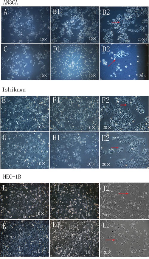
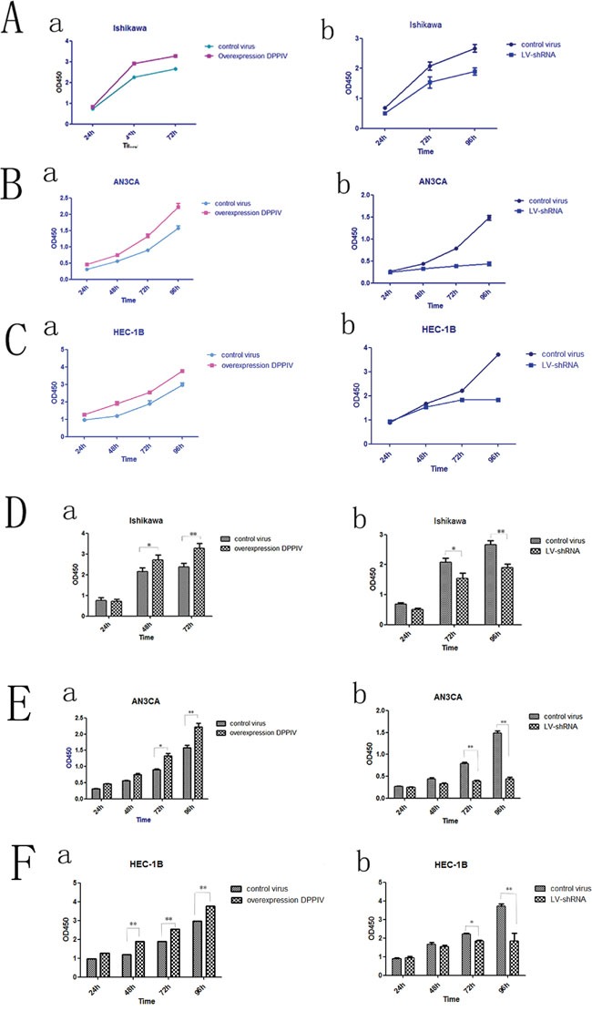
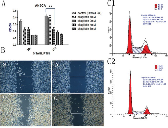
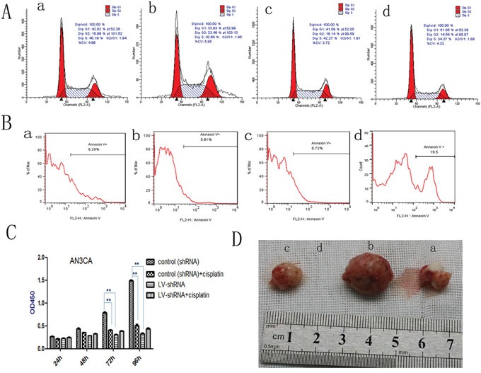
Dipeptidyl peptidase IV (DPPIV), also known as CD26, is a 110-kDa cell surface glycoprotein expressed in various tissues. DPPIV reportedly plays a direct role in the progression of several human malignancies. DPPIV specific inhibitors are employed as antidiabetics and could potentially be repurposed to enhance anti-tumor immunotherapies. In the present study, we investigated the correlation between DPPIV expression and tumor progression in endometrial carcinoma (EC). DPPIV overexpression altered cell morphology and stimulated cell proliferation, invasion and tumorigenesis in vitro and in vivo. These effects were abrogated by DPPIV knockdown or pharmacological inhibition using sitagliptin. DPPIV overexpression increased hypoxia-inducible factor 1a (HIF-1a) and vascular endothelial growth factor A (VEGFA) expression to promote HIF-1a-VEGFA signaling. Our results indicated that DPPIV accelerated endometrial carcinoma progression and that sitagliptin may be an effective anti-EC therapeutic.

Keywords: dipeptidyl peptidase IV; endometrial carcinoma; hypoxia-inducible factor 1a; sitagliptin; vascular endothelial growth factor A.


Conflict of interest statement

CONFLICTS OF INTEREST

The authors declare no conflicts of interest.

Figures

Figure 1
Figure 1. Morphological changes in Ishikawa, HEC-1B and AN3CA cells following DPPIV overexpression or knockdown
A–L. Cells overexpressing DPPIV were actively growing and had more pseudopodia-like connections between cells (arrow) after transfection for 96 h and first passage 24 h later (B1 and B2, AN3CA cells; F1 and F2, Ishikawa cells; J1 and J2, HEC-1B cells). ShRNA-mediated DDPIV knockdown altered cell morphology; filamentous connections between cells were lost and cells became apoptotic (arrow) (D1 and D2, AN3CA cells; H1 and H2, Ishikawa cells; L1 and L2, HEC-1B cells); A, C, E, G, I and K negative controls). (Cell morphology was recorded using an IX71 microscopy system coupled to a DP73 digital camera).
Figure 2
Figure 2. DPPIV overexpression increased cell proliferation in Ishikawa, HEC-1B and AN3CA cells, while shRNA-mediated DPPIV knockdown had the opposite effect
A-C. DPPIV overexpression stimulates proliferation while DPPIV knockdown suppressed growth in Ishikawa (A), AN3CA (B) and HEC-1B (C) cells. D-F. Quantitative analysis of results shown in A-C results represent mean ± SD (n = 4). *P < 0.05; **P < 0.01 (Student's t test).
Figure 5
Figure 5. DPPIV inhibitor- sitagliptin suppresses cell proliferation, cell migration and cell cycle
A. Cell viability is diminished by DDPIV inhibition. AN3CA cells were treated with the different concentrations DPPIV inhibitor-sitagliptin or left untreated and viability was evaluated 48 h later. Results are mean values ± SD (n = 4). **P < 0.001 (Student's t test). B. Cell migration in the absence (control) or presence of sitagliptin (1 mM). The graph is representative of three independent experiments. C. Cycle arrest at G0/G1 in the presence of sitagliptin (1 mM) (C2) or absence sitagliptin (C1).
Figure 3
Figure 3. Analysis of migration in DPPIV-overexpressing or knockdown cells
A–H. Ishikawa, HEC-1B and AN3CA cells were analyzed with the wound-healing assay at 0 h (A–D) and 24–48 h (E–H) after insert removal. A and E, overexpression control; B and F, DDPIV overexpression; C and G, shRNA control; D and H, LV-shRNA. Th migratory capacity of EC cells was enhanced by DPPIV overexpression (indicated by a line in F) and reduced by DPPIV knockdown (indicated by a line in H) especially in AN3CA cells after 24–48 h of culture.
Figure 4
Figure 4. DPPIV inhibitor induces cell cycle arrest, induces apoptosis; DPPIV knockdown and the chemotherapy sensitivity test; DPPIV overexpression increases tumorigenicity
A. DPPIV depletion induces cell cycle arrest at G0/G1, while DPPIV overexpression increases cells enter into S and G2 phase. B. DPPIV depletion induces apoptosis (B) in AN3CA cells, as determined by flow cytometry. C. DPPIV knockdown inhibited cell proliferation, similar to the effects of cisplatin. **P < 0.001, there were no synergistic effects associated with DPPIV knockdown and concurrent cisplatin treatment after 48, 72 and 96h. (P >0.05). The 95% confidence interval is (-0.887, -0.00129), (-0.960, -0.684), (-0.1560, -0.07248) respectively. D. DPPIV overexpression and knockdown compare with they control group at 8 weeks after injection. a, overexpression control; b, DDPIV overexpression; c, shRNA control; d, LV-shRNA. The graph is representative of three experiments.
Figure 6
Figure 6. In AN3CA cells, DPPIV overexpression increase the protien and mRNA expression of HIF-1α and VEGFA; DPPIV overexpression inceases the mRNA expression of IGF-1, but not IGF-1R
A. AN3CA cells after transfecte the vetor 96h, tained with anti VEGFA(1:400 ab51745, abcam) antibody, cell nucleus were stained with DAPI. Stained cells were viewed and imaged with immunofluorescence microscopy. a is the control virus-for overexpress; b is the overexpression DPPIV group; c is the control virus-for shRNA and d is the lentivirus-shRNA group (LV-shRNA). a1-d1 indicate nucleus stain; a2-d2 indicate cytoplasm stain; a-d is the merge. B, C. VEGFA protien level was tested by western blot(WB) and quantify the protien expression levels of these four groups. D, E. HIF-1α protien level in the four groups was tested by western blot(WB) and analysis. F-L. VEGFA, HIF-1α, IGF-1 and IGF-1R mRNA level in the four groups was tested by Quantitative real-time RT-PCR analysis. Over expression DPPIV increase the VEGFA, HIF-1α, IGF-1 mRNA expression compare with the control group, but there is no obvious change in IGF-1R genes. Results are mean values ± SD (n = 4). (Student's t-test, (*p<0.05; **p<0.001). The graph is representative of three independent experiments.

References

    1. Harkenrider MM, Block AM, Siddiqui ZA, Small W., Jr The role of vaginal cuff brachytherapy in endometrial cancer. Gynecologic oncology. 2015;136:365–372. - PubMed
    1. Jin F, Devesa SS, Zheng W, Blot WJ, Fraumeni JF, Jr, Gao YT. Cancer incidence trends in urban Shanghai, 1972-1989. International journal of cancer. 1993;53:764–770. - PubMed
    1. Ushijima K. Current status of gynecologic cancer in Japan. Journal of gynecologic oncology. 2009;20:67–71. - PMC - PubMed
    1. Ioffe YJ, Chiappinelli KB, Mutch DG, Zighelboim I, Goodfellow PJ. Phosphatase and tensin homolog (PTEN) pseudogene expression in endometrial cancer: a conserved regulatory mechanism important in tumorigenesis? Gynecologic oncology. 2012;124:340–346. - PMC - PubMed
    1. Fader AN, Arriba LN, Frasure HE, von Gruenigen VE. Endometrial cancer and obesity: epidemiology, biomarkers, prevention and survivorship. Gynecologic oncology. 2009;114:121–127. - PubMed

MeSH terms

Substances

LinkOut - more resources