. 2017 Feb 14;8(7):11963-11976.
doi: 10.18632/oncotarget.14449.

Comparison of the tumor cell secretome and patient sera for an accurate serum-based diagnosis of pancreatic ductal adenocarcinoma

Affiliations

Comparison of the tumor cell secretome and patient sera for an accurate serum-based diagnosis of pancreatic ductal adenocarcinoma

Shakhawan Mustafa et al. Oncotarget. .

Abstract

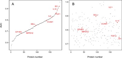
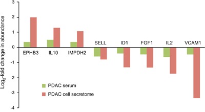
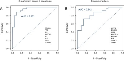
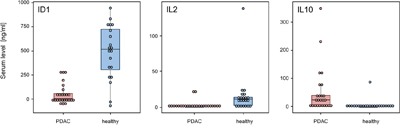
Pancreatic cancer is the currently most lethal malignancy. Toward an accurate diagnosis of the disease in body liquids, we studied the protein composition of the secretomes of 16 primary and established cell lines of pancreatic ductal adenocarcinoma (PDAC). Compared to the secretome of non-tumorous cells, 112 proteins exhibited significantly different abundances. Functionally, the proteins were associated with PDAC features, such as decreased apoptosis, better cell survival and immune cell regulation. The result was compared to profiles obtained from 164 serum samples from two independent cohorts - a training and a test set - of patients with PDAC or chronic pancreatitis and healthy donors. Eight of the 112 secretome proteins exhibited similar variations in their abundance in the serum profile specific for PDAC patients, which was composed of altogether 189 proteins. The 8 markers shared by secretome and serum yielded a 95.1% accuracy of distinguishing PDAC from healthy in a Receiver Operating Characteristic curve analysis, while any number of serum-only markers produced substantially less accurate results. Utility of the identified markers was confirmed by classical enzyme linked immunosorbent assays (ELISAs). The study highlights the value of cell secretome analysis as a means of defining reliable serum biomarkers.

Keywords: antibody microarray; biomarkers; cell lines; pancreatic cancer; secretome.


Conflict of interest statement

CONFLICTS OF INTEREST

These authors declare no conflicts of interest.

Figures

Figure 1
Figure 1. Proteins that are commonly regulated in secretome and intracellular proteome of tumor cells
The proteins are listed, which exhibited similar regulation in both secretome and proteome analyses of the ten established pancreatic cancer cell lines in comparison to non-tumor cells. The bar sizes indicate the relative degree of regulation.
Figure 2
Figure 2. AUC values of the 189 individual serum markers
Analysis by Receiver Operating Characteristic (ROC) curves was performed for all identified serum protein markers individually. Panel A shows the result calculated from the training set; the respective AUC values are shown, ranging from 55.2% to 96.0%. In panel B, the AUC values are shown as calculated for the individual marker molecules in the test set. For presentation, the order of the markers along the x-axis was kept as in panel A, highlighting the limited degree of reproducibility for individual markers.
Figure 3
Figure 3. Proteins that were similarly regulated in both tumor cell secretome and PDAC patient sera
Of the 112 differentially expressed proteins in the tumor cell secretome compared to the non-tumorous cells, only 8 were similarly regulated in the serum. The red and green bars indicate the degree of regulation in secretome and PDAC serum, respectively.
Figure 4
Figure 4. Diagnostic potential of the protein signatures in serum
A. A Receiver Operating Characteristic (ROC) curves was calculated for the signature of 8 proteins that exhibited shared expression changes in the analyses of PDAC serum and secretome, yielding an AUC value of 95.1%. The protein names are shown. B. In comparison, the ROC curve is presented for the best performing panel of 8 proteins that differed in abundance in the serum only; AUC is 84.2%. The relevant proteins are listed.
Figure 5
Figure 5. ELISA validation of identified marker proteins
The protein content was analyzed of 25 and 21 sera, respectively, isolated from PDAC patients and healthy blood donors. Proteins ID1 (A), IL2 (B), and IL10 (C) were studied with commercially available assays. All markers showed a highly significant degree of variation of abundance, with p-values < 0.001.

References

    1. Edwards BK, Noone AM, Mariotto AB, Simard EP, Boscoe FP, Henley SJ, Jemal A, Cho H, Anderson RN, Kohler BA, Eheman CR, Ward EM. Annual Report to the Nation on the status of cancer, 1975-2010, featuring prevalence of comorbidity and impact on survival among persons with lung, colorectal, breast, or prostate cancer. Cancer. 2014;120:1290–314. doi: 10.1002/cncr.28509. - DOI - PMC - PubMed
    1. Ferlay J, Steliarova-Foucher E, Lortet-Tieulent J, Rosso S, Coebergh JW, Comber H, Forman D, Bray F. Cancer incidence and mortality patterns in Europe: estimates for 40 countries in 2012. Eur J Cancer. 2013;49:1374–403. doi: 10.1016/j.ejca.2012.12.027. - DOI - PubMed
    1. Siegel RL, Miller KD, Jemal A. Cancer statistics, 2016. CA Cancer J Clin. 2016;66:7–30. doi: 10.3322/caac.21332. - DOI - PubMed
    1. Luo J, Xiao L, Wu C, Zheng Y, Zhao N. The incidence and survival rate of population-based pancreatic cancer patients: Shanghai cancer registry 2004-2009. PLOS ONE. 2013;8:e76052. doi: 10.1371/journal.pone.0076052. - DOI - PMC - PubMed
    1. Poruk KE, Firpo MA, Adler DG, Mulvihill SJ. Screening for pancreatic cancer: why, how, and who? Ann Surg. 2013;257:17–26. doi: 10.1097/SLA.0b013e31825ffbfb. - DOI - PMC - PubMed

Substances