. 2017 Jan 6;13(1):e1006537.
doi: 10.1371/journal.pgen.1006537. eCollection 2017 Jan.

Genomes of Fasciola hepatica from the Americas Reveal Colonization with Neorickettsia Endobacteria Related to the Agents of Potomac Horse and Human Sennetsu Fevers

Affiliations

Genomes of Fasciola hepatica from the Americas Reveal Colonization with Neorickettsia Endobacteria Related to the Agents of Potomac Horse and Human Sennetsu Fevers

Samantha N McNulty et al. PLoS Genet. .

Abstract

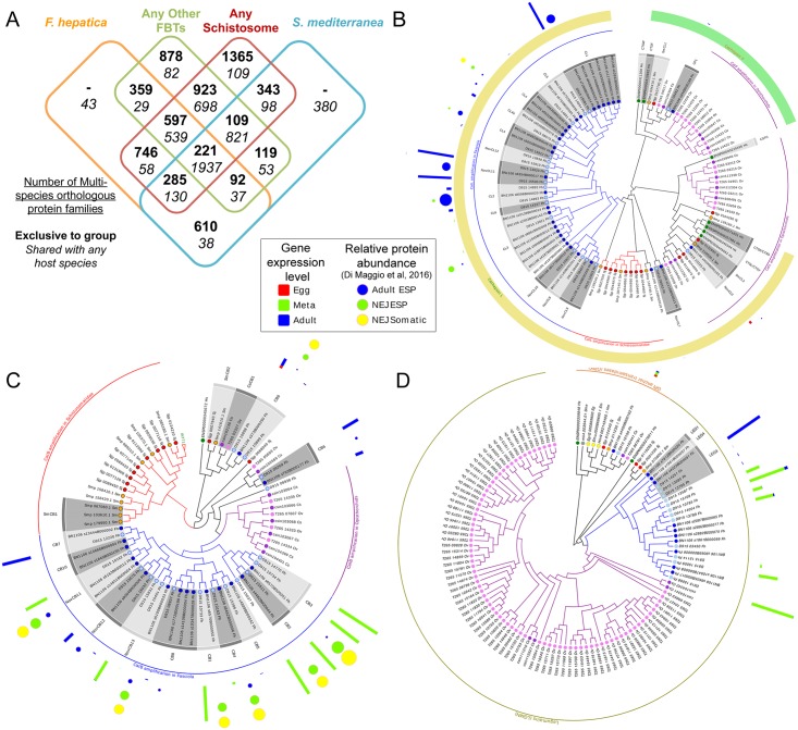
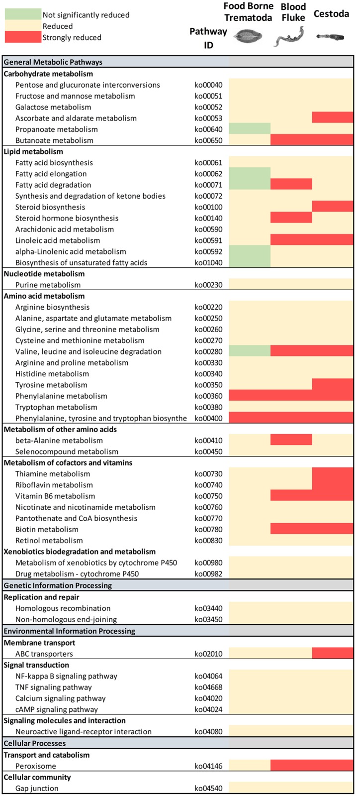
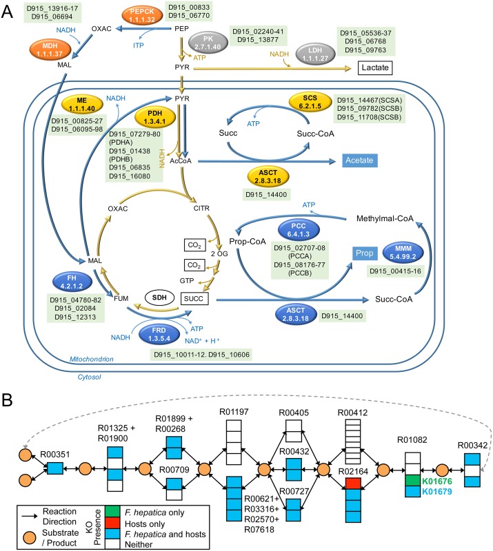
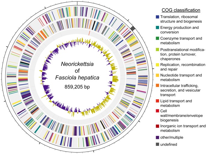
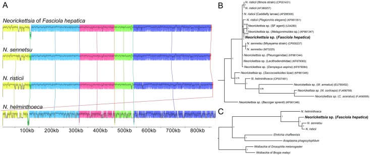
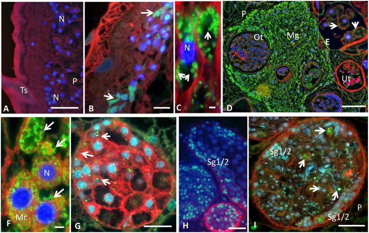
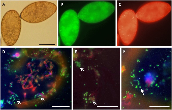
Food borne trematodes (FBTs) are an assemblage of platyhelminth parasites transmitted through the food chain, four of which are recognized as neglected tropical diseases (NTDs). Fascioliasis stands out among the other NTDs due to its broad and significant impact on both human and animal health, as Fasciola sp., are also considered major pathogens of domesticated ruminants. Here we present a reference genome sequence of the common liver fluke, Fasciola hepatica isolated from sheep, complementing previously reported isolate from cattle. A total of 14,642 genes were predicted from the 1.14 GB genome of the liver fluke. Comparative genomics indicated that F. hepatica Oregon and related food-borne trematodes are metabolically less constrained than schistosomes and cestodes, taking advantage of the richer millieux offered by the hepatobiliary organs. Protease families differentially expanded between diverse trematodes may facilitate migration and survival within the heterogeneous environments and niches within the mammalian host. Surprisingly, the sequencing of Oregon and Uruguay F. hepatica isolates led to the first discovery of an endobacteria in this species. Two contigs from the F. hepatica Oregon assembly were joined to complete the 859,205 bp genome of a novel Neorickettsia endobacterium (nFh) closely related to the etiological agents of human Sennetsu and Potomac horse fevers. Immunohistochemical studies targeting a Neorickettsia surface protein found nFh in specific organs and tissues of the adult trematode including the female reproductive tract, eggs, the Mehlis' gland, seminal vesicle, and oral suckers, suggesting putative routes for fluke-to-fluke and fluke-to-host transmission. The genomes of F. hepatica and nFh will serve as a resource for further exploration of the biology of F. hepatica, and specifically its newly discovered trans-kingdom interaction with nFh and the impact of both species on disease in ruminants and humans.


Conflict of interest statement

The authors have declared that no competing interests exist.

Figures

Fig 1
Fig 1. Protein families in trematodes.
(A) A Venn diagram demonstrating the phylogenetic distribution of orthologous protein families among the trematode species analyzed (B) Differential amplification of cathepsins L and F in trematodes. A maximum likelihood tree of the genes annotated as members of the C1A protease family from trematodes shows that while a single set of cathepsins F are detected in Schistosomes and F. hepatica strains, an expansion of cathepsins F is observed in O. viverrini and C. sinensis (green arch). Similarly, the cathepsins L-like of the trematodes (yellow arch) show a basal node more related to the mammalian enzymes and several independent amplifications in schistosomes, opistorchiids and Fasciola. Most known cathepsins variants are supported by expression data in different stages (red, green and blue bars), and proteomic data (yellow, green and blue dots) from a recent report [34]. Several putative novel variants are indicated, most of them not expressed at the stages analyzed. (C) Cathepsin B subfamily of the C1A protease family. A basal cathepsin B node and differential expansions in schistosomatids (red arch), fasciolids (blue arch) and opisthorchiids (purple arch) is observed. As in cathepsins L novel isoforms are identified, some of them supported by expression data. (D) Legumain is differentially amplified in food borne trematodes. Maximum likelihood tree of the genes annotated as members of the C13 protease family. While a single gene corresponding to the glycosyl-phosphatidyl-inositol anchor transamidase exists in all trematodes, amplification of the legumains was evident in the food borne trematodes F. hepatica, C. sinensis and O. viverrini, but not in the blood flukes S. mansoni and S. japonicum. These amplification events are independent in the different lineages. Species included in the tree are color coded (human, emerald dots, planaria (grey) cestodes (yellow) S. japonicum (dark red) S. mansoni (orange) C. sinensis (purple) O. viverrini (pink), F. hepatica Oregon strain (sky blue) F. hepatica Liverpool strain (navy).
Fig 2
Fig 2. Metabolic pathway reduction in parasitic flatworms.
The global number of proteins assigned to different metabolic pathway was compared between different parasitic flatworms, the free-living planaria and cattle predicted proteomes. Those pathways showing a significant reduction in the parasitic species in relation to those present in planaria are indicated (strongly reduced: less than 30% conservation; reduced: conservation between 30 and 80%; not significantly reduced: more than 80% conservation). While in general several pathways are reduced in all parasitic species, some pathways are differential between food borne trematodes (F. hepatica, O. viverrini, C. sinensis), blood flukes (S. mansoni and S. japonicum) and cestodes (E. multilocularis, H. microstoma and T. solium). Most notably some lipid metabolism and amino acid pathways (i.e. aliphatic amino acid degradation) are not reduced in FBT (green) while they are reduced in the other groups. In general, FBT seem to be less constrained than blood flukes, with cestodes being the most restricted metabolically.
Fig 3
Fig 3. Metabolic pathways in F. hepatica.
(A) Energy metabolism in anaerobic mitochondria of F. hepatica by malate dismutase. While the classical anaerobic fermentation to lactate is present (enzymes indicated in gray) when oxygen tension is low, the malate dismutation pathway is preferred (blue arrows). Phosphenol pyruvate reduction to malate occurs in the cytoplasm (orange). Within the mitochondria, part of the malate is oxidized to acetate (yellow) while other fraction reduced to succinate and further transformed to propionate (blue). Genes predicted for key enzymes involved in anaerobic respiration are indicated. Abbreviations: PEP, phosphenolpyruvate; OXAC, oxaloacetate; MAL, malate; FUM, fumarate; SUCC, succinate; PYR, pyruvate; AcCoA, acetyl-CoA; CITR, citrate. Enzymes indicated are: PK, pyruvate kinase; LDH, lactate dehydrogenase, PEPCK, phosphenolpyruvate carboxykinase (ATP dependent); MDH, malate dehydrogenase; ME, malic enzyme; PDH, pyruvate dehydrogenase; ASCT, acetate:succinate CoA-transferase; SCS, succinyl-CoA synthetase; FH, fumarate hydratase; FRD, fumarate reductase; SDH, succinate dehydrogenase, MMM methylmalonyl-CoA mutase; PCC, propionyl-CoA carboxylase. (B) Parasite specific enzyme usage in TCA cycle. The KEGG module for TCA cycle (M00009) is shown with groups of orthologous enzymes indicated using KEGG orthology (KO) IDs. An interesting example of alternate enzyme usage is shown for fumarate hydratase (reaction R01082), catalyzed by a class II enzyme (EC 4.2.1.2B; K01679) in both the host and parasite. However, F. hepatica also has a Platyhelminthes-specific class I fumarate hydratase (EC 4.2.1.2A; K01676), not annotated in any of the host proteomes. Such knowledge can be leveraged to design worm specific therapies with potentially low (or no) impact on the host health.
Fig 4
Fig 4. The genome of the Neorickettsia endobacterium of Fasciola hepatica.
The first track (from outside to inside) represents the 859,205bp genome of the Neorickettsia endobacterium of Fasciola hepatica (100 kb major ticks, 10 kb minor ticks). The genome shows nearly complete synteny with Neorickettsia risticii and Neorickettsia sennetsu with the exception of an inversion (shaded in grey). The second and third tracks represent the 744 inferred protein coding genes on the plus and minus strands, respectively. Genes are coded based on their NCBI Clusters of Orthologous Groups of proteins database classification. The fourth track represents RNA coding genes, including 3 ribosomal (red), 33 transfer (black), and 1 short, noncoding (blue). The fifth track depicts the G-C skew [(G-C)/(G+C)] calculated over 500bp windows.
Fig 5
Fig 5. Phylogenetic affinities of the Neorickettsia symbiont of Fasciola hepatica.
(A) The genomes of the four fully sequenced Neorickettsia species were aligned. Syntenic blocks are colored. The genome of the Neorickettsia of Fasciola hepatica (PRJNA295290) shares almost complete synteny with the genomes of Neorickettsia risticii (PRJNA19099) and Neorickettsia sennetsu (PRJNA357), with the exception of a small inversion that is also present in Neorickettsia helminthoeca (PRJNA187358). (B) Bayesian inference phylogenetic analysis based on available 16S ribosomal RNA sequences of Neorickettsia species, retrieved from [15]. While the resolution of the sub-clade consisting of Neorickettsia risticii and Neorickettsia sennetsu is sub-optimal, the tree indicates that the Neorickettsia of Fasciola hepatica (nFh) may be most closely related to a strain that occurs in Metagonimoides species and an agent of Sennetsu fever. (C) A Bayesian inference phylogenetic tree based on the protein sequences of 473 single-copy, orthologous protein families conserved in the represented species clearly indicates that nFh is more closely related to N. risticii and N. sennetsu than N. helminthoeca or other species of the family Anaplasmataceae. NCBI GenBank accession numbers are indicated. Trematode hosts or other defining features are indicated for uncharacterized species.
Fig 6
Fig 6. Immunofluorescence detection of Neorickettsia in adult Fasciola hepatica using polyclonal anti-serum raised against a recombinant surface protein of Neorickettsia of P. elegans (PeNsp-3, green labeling).
DAPI (blue) and wheat hemagglutinin (red) were used to detect double stranded DNA and plasma membranes, respectively. (A) No green labeling was seen in the tegument of F. hepatica from Uruguay that were known to be devoid of Neorickettsia. (B) Clusters of Neorickettsia (arrows) in the tegument close to tegumental nuclei in F. hepatica from Oregon. (C) Numerous ‘donut’-shaped endobacteria (arrows) in the parenchyma in F. hepatica from Oregon. (D) Labeling of large numbers of Neorickettsia in the Mehlis’ gland and labeling of single endobacterium in the ootype or intrauterine eggs of F. hepatica from Oregon. (E) Magnification of a region proximal to (D) showing granular staining of single endobacterium in intrauterine eggs. (F) Clusters of Neorickettsia rods (arrows) in the cytoplasm of Mehlis’ cells of F. hepatica Oregon. (G) Individual Neorickettsia endobacteria (arrows) in a vitelline follicle with different stages of vitelline cells of F. hepatica Oregon. (H) No green staining indicative of Neorickettsia were found in the testis of F. hepatica from Uruguay tested negative for Neorickettsia by PCR. (I) Neorickettsia endobacteria (arrows) in the testis of F. hepatica Oregon with spermatogonia in the periphery and developing spermatozoa in the center. N, nucleus; Ts, tegument spine; P, parenchyma; Mg, Mehlis’ gland; Ot, ootype; Ut, uterus; Mc, Mehlis’ cell; Sg1/2, primary and secondary spermatogonia; Bar corresponds in A-B, D-I to 100 μm and in C to 1 μm.
Fig 7
Fig 7. Immunofluorescence detection of Neorickettsia in eggs of F. hepatica from Oregon using polyclonal anti-serum raised against a recombinant surface protein of Neorickettsia of P. elegans (PeNsp-3, green labeling, D-F).
(A) Unstained eggs recovered from the liver by regular light microcopy. (B) and (C) Unstained eggs recovered from the liver by immunofluorescence microscopy using different filters demonstrating auto-fluorescence. (D-F) Cross-sections of eggs showing various amounts of Neorickettsia (arrows). Bar corresponds to 50 μm.

References

    1. Keiser J, Utzinger J. Food-borne trematodiases. Clin Microbiol Rev. 2009;22(3):466–83. 10.1128/CMR.00012-09 - DOI - PMC - PubMed
    1. Hotez PJ, Alvarado M, Basanez MG, Bolliger I, Bourne R, Boussinesq M, et al. The global burden of disease study 2010: interpretation and implications for the neglected tropical diseases. PLoS Negl Trop Dis. 2014;8(7):e2865 10.1371/journal.pntd.0002865 - DOI - PMC - PubMed
    1. Torgerson PR, Devleesschauwer B, Praet N, Speybroeck N, Willingham AL, Kasuga F, et al. World Health Organization Estimates of the Global and Regional Disease Burden of 11 Foodborne Parasitic Diseases, 2010: A Data Synthesis. PLoS Med. 2015;12(12):e1001920 10.1371/journal.pmed.1001920 - DOI - PMC - PubMed
    1. Nyindo M, Lukambagire AH. Fascioliasis: An Ongoing Zoonotic Trematode Infection. Biomed Res Int. 2015;2015:786195 10.1155/2015/786195 - DOI - PMC - PubMed
    1. Charlier J, Vercruysse J, Morgan E, van Dijk J, Williams DJ. Recent advances in the diagnosis, impact on production and prediction of Fasciola hepatica in cattle. Parasitology. 2014;141(3):326–35. Epub 2013/11/16. 10.1017/S0031182013001662 - DOI - PubMed

Publication types

MeSH terms

Substances

Supplementary concepts

LinkOut - more resources