. 2017 Jan 6;12(1):e0168059.
doi: 10.1371/journal.pone.0168059. eCollection 2017.

Enhanced Chondrogenic Differentiation of Human Umbilical Cord Wharton's Jelly Derived Mesenchymal Stem Cells by GSK-3 Inhibitors

Affiliations

Enhanced Chondrogenic Differentiation of Human Umbilical Cord Wharton's Jelly Derived Mesenchymal Stem Cells by GSK-3 Inhibitors

Prapot Tanthaisong et al. PLoS One. .

Abstract

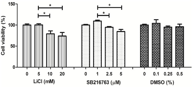
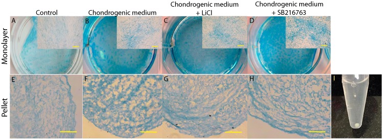
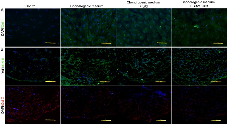
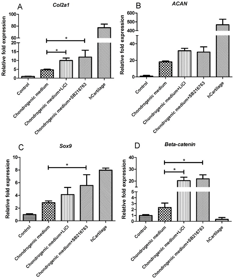
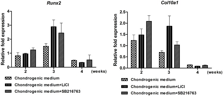
Articular cartilage is an avascular, alymphatic, and aneural system with very low regeneration potential because of its limited capacity for self-repair. Mesenchymal stem cells (MSCs) are the preferred choice for cell-based therapies. Glycogen synthase kinase 3 (GSK-3) inhibitors are compounds that can induce the Wnt signaling pathway, which is involved in chondrogenesis and cartilage development. Here, we investigated the influence of lithium chloride (LiCl) and SB216763 synergistically with TGF-β3 on chondrogenic differentiation in human mesenchymal stem cells derived from Wharton's jelly tissue (hWJ-MSCs). hWJ-MSCs were cultured and chondrogenic differentiation was induced in monolayer and pellet experiments using chondrogenic medium, chondrogenic medium supplemented with LiCl, or SB216763 for 4 weeks. After in vitro differentiation, cultured cells were examined for the expression of Sox9, ACAN, Col2a1, and β-catenin markers. Glycosaminoglycan (GAG) accumulation was also examined by Alcian blue staining. The results indicated that SB216763 was more effective than LiCl as evidenced by a higher up-regulation of the expression of cartilage-specific markers, including Sox9, ACAN, Col2a1 as well as GAG accumulation. Moreover, collagen type II expression was strongly observed in cells cultured in the chondrogenic medium + SB216763 as evidenced by western blot analysis. Both treatments appeared to mediate the Wnt signaling pathway by up-regulating β-catenin gene expression. Further analyses showed that all treatments suppressed the progression of chondrocyte hypertrophy, determined by decreased expression of Col10a1 and Runx2. These results indicate that LiCl and SB216763 are potential candidates for further in vivo therapeutic trials and would be of great importance for cartilage regeneration.


Conflict of interest statement

This study was partly supported by the Bangkok Stem Cell Co., Ltd. There are no patents, products in development or marketed products to declare. This does not alter our adherence to all the PLOS ONE policies on sharing data and materials, as detailed online in the guide for authors.

Figures

Fig 1
Fig 1. Morphology of hWJ-MSCs with a typical fibroblast-like morphology.
(A) Phase contrast images of hWJ-MSCs expanded from Wharton’s jelly tissue (arrow) and (B) hWJ-MSCs at 80% confluences. Scale bar = 10 μm.
Fig 2
Fig 2. Characterization of hWJ-MSCs.
(A) Immunophenotype of MSCs, immunofluorescent micrographs staining expression of MSC markers (CD73, 90, and 105), Nuclei were counterstains with DAPI (blue). Cells were negative for hematopoietic marker (CD34). Scale bar = 20 μm. (B) Differentiation of hWJ-MSCs to mesodermal linage cells. The cells were induced to undergo adipogenic, osteogenic, and condrogenic differentiation.
Fig 3
Fig 3. The toxicity effect of LiCl and SB216763 on hWJ-MSC viability.
hWJ-MSCs were cultured with 0–20 mM LiCl (A), 0–5 μM SB216763 (B), or 0–0.5% DMSO (C), for 72 hrs in 96-well plate. Then, the viability was detected by MTT assay. **DMSO without chemical was use as vehicle control. **Data were exposed as mean ± SD. *P<0.05.
Fig 4
Fig 4. Accumulation of GAGs was stained by alcian blue.
(A-D) Photographs of monolayer expanded cells cultured for 2 weeks. Scale bar = 10 μm. (E-H) Pellets culture at 4 weeks after differentiation. Scale bar = 20 μm. (I) Morphology of pellet culture at 4 weeks after differentiation.
Fig 5
Fig 5. Immunofluorescent staining of cartilage specific type collagen.
(A) immunofluorescent staining for collagen type II in monolayer expanded cultured on 3 weeks after differentiation. Scale bar = 20 μm. (B) Collagen type II and X expressions in pellet experiment on 4 weeks after differentiation. Scale bar = 20 μm.
Fig 6
Fig 6. The collagen type II protein after 4 weeks of inductions examined by western blot analysis.
β-actin was used as an internal control.
Fig 7
Fig 7. qPCR analysis for chondrogenic gene expressions after 4 weeks of inductions.
(A) Col2a1, (B) ACAN, (C) Sox9 and (D) β-catenin. Gene expression was normalized to coresponding GAPDH and calculated by relative expression compared to control cells. The experiments were perfromed three times. **Data were expressed as mean±SD, *P<0.05.
Fig 8
Fig 8. Expression level of hypertrophic marker genes (A) Col10a1, and (B) Runx2 was quantified by qPCR after 2, 3, and 4 weeks of inductions.
Gene expression was normalized to coresponding GAPDH and calculated by relative expression compared to control cells. Data were expressed as mean±SD, The expperiments were perfromed three times.

References

    1. Fox AS, Bedi A, Rodeo SA. The basic science of articular cartilage: structure, composition, and function. Sports Health. 2009; 1: 461–468. 10.1177/1941738109350438 - DOI - PMC - PubMed
    1. Pittenger MF, Mackay AM, Beck SC, Jaiswal RK, Douglas R, Mosca JD, et al. Multilineage potential of adult human mesenchymal stem cells. Science. 1999; 284(5411): 143–147. - PubMed
    1. Zuk PA, Zhu M, Mizuno H, Huang J, Futrell JW, Katz AJ, et al. Multilineage cells from human adipose tissue: implications for cell-based therapies. Tis Eng. 2001; 7: 211–228. - PubMed
    1. Gronthos S, Mankani M, Brahim J, Robey PG, Shi S. Postnatal human dental pulp stem cells (DPSCs) in vitro and in vivo. Proc Natl Acad Sci U S A. 2000; 97: 13625–13630. 10.1073/pnas.240309797 - DOI - PMC - PubMed
    1. Phuc P, Nhung T, Loan D, Chung D, Ngoc P. Differentiating of banked human umbilical cord blood-derived mesenchymal stem cells into insulin-secreting cells. In Vitro Cell Dev Biol Anim. 2011; 47(1): 54–63. 10.1007/s11626-010-9356-5 - DOI - PubMed

MeSH terms