. 2017 Jan 6;12(1):e0169639.
doi: 10.1371/journal.pone.0169639. eCollection 2017.

Past1 Modulates Drosophila Eye Development

Affiliations

Past1 Modulates Drosophila Eye Development

Orly Dorot et al. PLoS One. .

Erratum in

Abstract

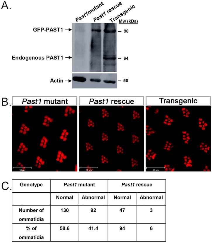
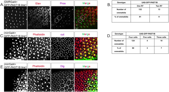
Endocytosis is a multi-step process involving a large number of proteins, both general factors, such as clathrin and adaptor protein complexes, and unique proteins, which modulate specialized endocytic processes, like the EHD proteins. EHDs are a family of Eps15 Homology Domain containing proteins that consists of four mammalian homologs, one C. elegans, one Drosophila melanogaster and two plants orthologs. These membrane-associated proteins are involved in different steps of endocytic trafficking pathways. We have previously shown that the Drosophila EHD ortholog, PAST1, associates predominantly with the plasma membrane. Mutations in Past1 result in defects in endocytosis, male sterility, temperature sensitivity and premature death of the flies. Also, Past1 genetically interacts with Notch. In the present study, we investigated the role of PAST1 in the developing fly eye. In mutant flies lacking PAST1, abnormal differentiation of photoreceptors R1, R6 and R7 was evident, with partial penetrance. Likewise, five cone cells were present instead of four. Expression of transgenic PAST1 resulted in a dominant negative effect, with a phenotype similar to that of the deletion mutant, and appearance of additional inter-ommatidial pigment cells. Our results strongly suggest a role for PAST1 in differentiation of photoreceptors R1/R6/R7 and cone cells of the fly ommatidia.


Conflict of interest statement

The authors have declared that no competing interests exist.

Figures

Fig 1
Fig 1. Past1 mutant flies contain an abnormal number of photoreceptors in their eyes.
(A) Schematic representation of the apical section of an ommatidium. (B) Schematic representation of a cross-section of an ommatidium. (C) Scanning electron microscopy images of eyes of wild type or homozygous null Past1 mutant (Past1110-1) three-days-old adult flies. Upper panels x240, lower panels x1500. (D) Transmission electron microscopy images of eyes of wild type or Past1110-1 three-days-old adult flies. Orange frame delineates an example of an ommatidium with fewer photoreceptors than in the wild type. Blue frame delineates an example of an ommatidium with two potential R7 photoreceptors.
Fig 2
Fig 2. Ommatidial defects in Past1 mutant flies.
(A) Phalloidin staining of wild type or Past1110-1 homozygous mutant late pupa eyes. Dotted squares mark ommatidia with less than seven rhabdomeres. (B) Quantification of abnormal ommatidia (with less than seven rhabdomeres) in late pupal eyes of wild type (n = 229) and of Past1110-1 mutants (n = 222) ommatidia; P < 0.001. (C) Elav (red) and chapotin (green) staining of wild type and Past1110-1mutant early-mid pupa eyes (42-48h after puparium formation). The dotted square outlines an ommatidium with eight normal photoreceptors in wild type and an ommatidium with abnormal seven photoreceptors in Past1110-1mutant eyes. (D) Quantification of abnormal number of photoreceptors in early-mid pupa eyes of wild type (n = 198) and of Past1110-1 mutant (n = 123) ommatidia; P < 0.001.
Fig 3
Fig 3. Photoreceptor differentiation in Past1 mutant fly eye.
(A) Senseless staining of wild type and Past1110-1 homozygous mutant eye discs from third instar larvae. Shown are a Z-projections of confocal sections. (B) Phalloidin (red) and rhodopsin 1 (Rh1) (grey) staining of wild type Past1110-1 homozygous mutant late pupal eyes (85-96h after puparium formation). A schematic illustration depicting the localization of different photoreceptors in each zoomed-in image is shown on the right of each panel: I—ommatidium with normal seven photoreceptors (R1-R7), II—photoreceptor R6 is missing, III—photoreceptor R1 is missing, IV—photoreceptor R6 is missing, V—photoreceptor R1 is missing and there are two photoreceptors R7. The dotted circles outline the missing R1/R6 photoreceptors.
Fig 4
Fig 4. Specification of R1/R6 and R7 photoreceptors in Past1 mutant eyes.
(A) BarH1 (grey, or red in the merge) and Elav (grey, or green in the merge) staining of wild type and Past1110-1 homozygous mutant eye discs from third instar larvae. Shown are Z-projections of confocal sections. The yellow arrow marks the missing photoreceptors. (B) Elav (grey, or red in the merge) and pros (grey, or green in the merge) staining of wild type and Past1110-1mutant early-mid pupa eyes (42-48h after puparium formation). The dotted line marks the equator of the eye, defining the dorsal and ventral parts of the eye. The dotted circle delineates an ommatidium with two R7 photoreceptors and the dotted rectangle outlines two neighboring ommatidia that face each other in the same dorsal part of the eye. (C) Quantification of ommatidia containing one or two R7 photoreceptor in wild type (n = 249) and Past1110-1mutant (n = 278) ommatidia; P < 0.001.
Fig 5
Fig 5. Specification of cone and inter-ommatidial cells in Past1 mutant eyes.
(A) Elav (grey, or red in the merge) and cut (grey, or green in the merge) staining of wild type and Past1110-1 homozygous mutant early-mid pupal eyes (42-48h after puparium formation). Shown are Z-projections of confocal sections. The enlarged images on the right of each frame show an ommatidium with four cone cells in wild type and an ommatidium with five cone cells in Past1110-1mutant eyes. (B) Quantification of four or five cone cells in wild type (n = 189) and Past1110-1mutant (n = 186) ommatidia; P < 0.001. (C) Dlg (green) staining of wild type and Past1110-1mutant early-mid pupal eyes (42-48h after puparium formation). Shown are Z-projections of confocal sections. The enlarged images on the right of each frame show an ommatidium with nine inter-ommatidial pigment cells and four cone cells in wild type and an ommatidium with nine inter-ommatidial pigment cells and five cone cells in Past1110-1mutant eyes. (D) Dlg (green) and phalloidin (red) staining of wild type and Past1110-1mutant late pupal eyes (85-96h after puparium formation). Different focal planes of the same ommatidium, presenting: upper sections of the ommatidia for Dlg and lower sections for phalloidin.
Fig 6
Fig 6. Rescue of ommatidial defects in Past1 mutant by PAST1B transgenic expression.
(A) Lysates of one day old adult fly heads with the following genotypes: 1) GMRGal4>Sco/CyO; Past1110-1/Past1110-1 (negative control), 2) GMRGal4>GFP-PAST1B/CyO; Past1110-1/Past1110-1 (rescue), 3) GMRGal4>GFP-PAST1B/CyO; Sb/TM6b (transgenic/GFP-PAST1), were subjected to SDS-PAGE. The corresponding blot was interacted with anti-PAST1 serum and anti-actin antibody. (B) Phalloidin (red) staining of homozygous Past1110-1, GMRGal4>GFP-PAST1B/CyO; Past1110-1/ Past1110-1 and GMRGal4>GFP-PAST1B/CyO; Sb/TM6b, late pupa eyes (85-96h after puparium formation). (C) Quantification of rhabdomeres number in late pupa eyes of Past1110-1 homozygous mutant (n = 222) and GMRGal4>GFP-PAST1B/CyO; Past1110-1/ Past1110-1 (rescue) (n = 50) ommatidia; P < 0.001flies.
Fig 7
Fig 7. Overexpression of PAST1 in the eyes results in fused ommatidia.
(A) Scanning electron microscopy images show eyes of three-days-old adult flies. Upper panels: GMRGal4 (control), GMRGal4>UAS-GFP-PAST1B/TM6B (line 2), GMRGal4>UAS-GFP-PAST1A/TM6B (line 2), GMRGal4>UAS-GFP-PAST1B/CyO (line 1) and GMRGal4>UAS-GFP-PAST1A/CyO (line 1). Lower panels: GMRGal4>UAS-GFP-PAST1B/UAS-GFP-PAST1B (line 2), GMRGal4>UAS-GFP-PAST1A/UAS-GFP-PAST1A (line 2), GMRGal4>UAS-GFP-PAST1B/UAS-GFP-PAST1B (line 1) and GMRGal4>UAS-GFP-PAST1A/UAS-GFP-PAST1A (line 1). Differences in bristle numbers were not taken into account since they also appear in the UAS-GFP control flies. (B) Lysates from GMRGal4>UAS-GFP-PAST1B/UAS-GFP-PAST1B (line 2), GMRGal4>UAS-GFP-PAST1A/UAS-GFP-PAST1A (line 2), GMRGal4>UAS-GFP-PAST1B/UAS-GFP-PAST1B (line 1), GMRGal4>UAS-GFP-PAST1A/UAS-GFP-PAST1A (line 1) and GMRGal4>UAS-GFP (control) adult flies were subjected to SDS-PAGE and western blotting using anti-PAST1 and anti-actin antibodies. Quantification of GFP-PAST1 expression is shown at the bottom of the western blotting. In each lane, the band intensity of GFP-PAST1 expression was divided by the intensity of endogenous PAST1 band, both detected using anti-PAST1 antibodies. The results are the mean of four different experiments. Stars denote fusion between ommatidia.
Fig 8
Fig 8. Transgenic GFP-PAST1B contains an abnormal number of photoreceptors.
(A) Phalloidin (grey, or red in the merge) staining of GMRGal4>UAS-GFP-PAST1B/CyO (line 1) or GMRGal4>UAS-GFP-PAST1B/TM6b (GFP appears as grey, or green in the merge) (line 2) late pupal eyes (85-96h after puparium formation). (B) Quantification of abnormal ommatidia (ommatidium with more or less seven rhabdomeres) in late pupal eyes of PAST1B (line 1) (n = 135) or PAST1B (line 2) (n = 115) ommatidia; P < 0.001 only for PAST1B (line 2).
Fig 9
Fig 9. Specification of R7, cone and inter-ommatidial pigment cells in GFP-PAST1B transgenic flies.
(A) Staining of GMRGal4>UAS-GFP-PAST1B (GFP appears as grey, or green in the merge, in all the panels) (line 1), early-mid pupa eyes (42-48h after puparium formation) with Elav (grey, or red in the merge) and pros (grey, or purple in the merge). The dotted circle represents an ommatidium with two R7 photoreceptors (B) Quantification of the number of R7 photoreceptors in early-mid pupa eyes of wild type (n = 249) and overexpressing transgenic PAST1B (line 1) (n = 145) ommatidium; P < 0.001. (C) Phalloidin (grey, or red in the merge) and cut (grey or purple in the merge) staining of mirrGal4>UAS-GFP-PAST1B (line 1) early-mid pupa eyes (42-48h after puparium formation). The dotted circles represent ommatidia with five cone cells and the dotted square represents an ommatidium with three cone cells. Shown are Z-projections of confocal sections. (D) Quantification of the number of cone cells in early-mid pupal eyes of wild type (n = 189) and transgenic PAST1B (line 1) (n = 138) ommatidia; P < 0.002. (E) Phalloidin (grey, or red in the merge) and Dlg (grey, or purple in the merge) staining of mirrGal4>UAS-GFP-PAST1B (line 1) early-mid pupa eyes (42-48h after puparium formation). The arrowhead marks an extra inter-ommatidial pigment cell.
Fig 10
Fig 10. Overexpression of PAST1 attenuates endocytosis in Garland cells.
(A) Garland cells from third instar larvae of the following genotypes were dissected and incubated with Texas-red-conjugated avidin for one minute following 20 minutes of chase, fixed and visualized: DaGal4>UAS-GFP (control), DaGal4>UAS-GFP-PAST1A (line 1), DaGal4>UAS-GFP-PAST1B (line 1), DaGal4>UAS-GFP-PAST1A (line 2) and DaGal4>UAS-GFP-PAST1B (line 2). Cells were classified into: H—high level of internalization of avidin (wild type level of endocytosis), L—low level of internalization of avidin (reduced or no endocytosis), N—no internalization of avidin. (B) Quantification of avidin intensity in Garland cells of DaGal4>UAS-GFP (control) (n = 77), DaGal4>UAS-GFP-PAST1A (line 1) (n = 106), P < 0.002 and DaGal4>UAS-GFP-PAST1B (line 1) (n = 76); P < 0.001.
Fig 11
Fig 11. A model for ommatidium development.
(A) According to Tomlinson et al. 2011, expression of Notch in R1 or R6 leads to their development into R7 or cone cells. (B) A model for ommatidium development, presenting the impact of Past1 mutation (Past1null) on development of R1/R6/R7 and the cone cells. We assume that when overexpressed there is PAST1-mediated downregulation of endocytic processes, as shown in Fig 10, and therefore it behaves like a mutant Past1, that leads to Notch overexpression.

Similar articles

Cited by

References

    1. Drysdale RA, Crosby MA. FlyBase: genes and gene models. Nucleic Acids Res. 2005; 33 (Database issue): D390–5. 10.1093/nar/gki046 - DOI - PMC - PubMed
    1. Olswang-Kutz Y, Gertel Y, Benjamin S, Sela O, Pekar O, Arama E, et al. Drosophila Past1 is involved in endocytosis and is required for germline development and survival of the adult fly. J Cell Sci. 2009; 122: 471–80. 10.1242/jcs.038521 - DOI - PubMed
    1. Koles K, Messelaar EM, Feiger Z, Yu CJ, Frank CA, Rodal AA. The EHD protein Past1 controls postsynaptic membrane elaboration and synaptic function. Molecular Biology of the Cell. 2015; 26: 3275–88. 10.1091/mbc.E15-02-0093 - DOI - PMC - PubMed
    1. Fortini ME. Notch signaling: the core pathway and its posttranslational regulation. Dev Cell. 2009; 16: 633–47. 10.1016/j.devcel.2009.03.010 - DOI - PubMed
    1. Lorincz P, Takats S, Karpati M, Juhasz G. iFly: The eye of the fruit fly as a model to study autophagy and related trafficking pathways. Experimental Eye Research. 2016; 144: 90–8. 10.1016/j.exer.2015.06.013 - DOI - PubMed

LinkOut - more resources