. 2017 Oct;46(4):1167-1176.
doi: 10.1002/jmri.25602. Epub 2017 Jan 6.

Impact of vessel wall lesions and vascular stenoses on cerebrovascular reactivity in patients with intracranial stenotic disease

Affiliations

Impact of vessel wall lesions and vascular stenoses on cerebrovascular reactivity in patients with intracranial stenotic disease

Petrice M Cogswell et al. J Magn Reson Imaging. 2017 Oct.

Abstract

Purpose: To compare cerebrovascular reactivity (CVR) and CVR lagtimes in flow territories perfused by vessels with vs. without proximal arterial wall disease and/or stenosis, separately in patients with atherosclerotic and nonatherosclerotic (moyamoya) intracranial stenosis.

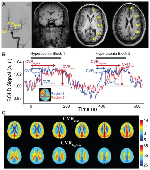
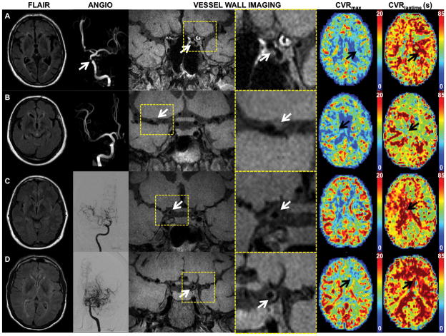
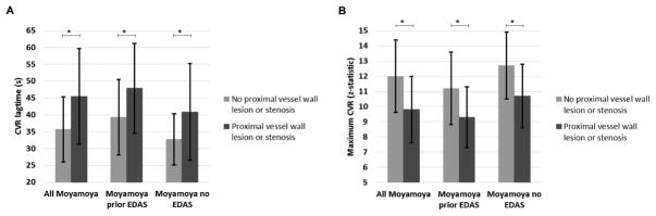
Materials and methods: Atherosclerotic and moyamoya patients with >50% intracranial stenosis and <70% cervical stenosis underwent angiography, vessel wall imaging (VWI), and CVR-weighted imaging (n = 36; vessel segments evaluated = 396). Angiography and VWI were evaluated for stenosis locations and vessel wall lesions. Maximum CVR and CVR lagtime were contrasted between vascular territories with and without proximal intracranial vessel wall lesions and stenosis, and a Wilcoxon rank-sum was test used to determine differences (criteria: corrected two-sided P < 0.05).

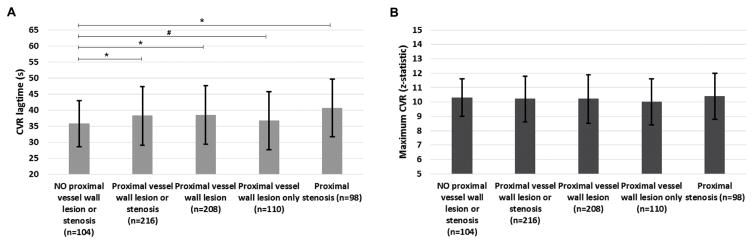
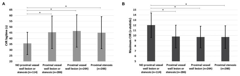
Results: CVR lagtime was prolonged in territories with vs. without a proximal vessel wall lesion or stenosis for both patient groups: moyamoya (CVR lagtime = 45.5 sec ± 14.2 sec vs. 35.7 sec ± 9.7 sec, P < 0.001) and atherosclerosis (CVR lagtime = 38.2 sec ± 9.1 sec vs. 35.0 sec ± 7.2 sec, P = 0.001). For reactivity, a significant decrease in maximum CVR in the moyamoya group only (maximum CVR = 9.8 ± 2.2 vs. 12.0 ± 2.4, P < 0.001) was observed.

Conclusion: Arterial vessel wall lesions detected on noninvasive, noncontrast intracranial VWI in patients with intracranial stenosis correlate on average with tissue-level impairment on CVR-weighted imaging.

Level of evidence: 4 Technical Efficacy: Stage 3 J. Magn. Reson. Imaging 2017;46:1167-1176.

Keywords: cerebrovascular reactivity; intracranial stenosis; moyamoya; vessel wall imaging.


Figures

Figure 1
Figure 1
(A) Representative images including: Digital subtraction angiography (DSA) with left internal carotid artery (ICA) injection showing the anterior territory segments for which degree of stenosis was graded, single coronal slice from vessel wall imaging exam, and T1-weighted images with the sub-territories labeled. (B) Blood-oxygenation-level-dependent (BOLD) cerebrovascular reactivity (CVR)-weighted determination. Time regression analysis is applied to record the time when the stimulus regressor generates the highest z-statistic with the data on a voxel-wise basis. The time shift is recorded as the CVR lagtime and the maximum z-statistic as the maximum CVR. (C) Mean CVR lagtime and maximum CVR values across all patients, which provide a reference standard for individual patients shown in Figure 2.
Figure 2
Figure 2
Axial FLAIR, MRA or DSA, coronal vessel wall imaging (subsection and magnified views), axial maximum CVR and CVR lagtime maps are shown for representative patients. (A) An 83 year male with atherosclerosis and left > right internal carotid artery (ICA) vessel wall lesions and stenosis (white arrows). Left middle cerebral artery (MCA) and anterior cerebral artery (ACA) territories (black arrows) show decreased maximum CVR (CVRmax) and prolonged CVR lagtime (CVRlagtime) compared to the right. (B) 71 year female with atherosclerosis. Vessel wall imaging demonstrates thickening and T1 hyperintensity of the proximal MCA (white arrow). No MCA stenosis is identified on MRA. CVR lagtime is prolonged in the right MCA territory (black arrow). A severe basilar stenosis may account for prolonged CVR lagtimes in the posterior territories bilaterally. (C) Vessel wall imaging in this 44 year female with predominantly right sided moyamoya disease is consistent with concentric thickening of the right terminal ICA (white arrow), proximal ACA, and proximal MCA. DSA right ICA injection shows moyamoya changes of the right anterior circulation. CVR lagtime is prolonged on the right (black arrow), but the maximum CVR is comparable to that on the left. (D) A 49 year female with bilateral, left>right, moyamoya disease and left > right vessel wall thickening of the terminal ICA (white arrow) and moyamoya changes. CVR lagtime is prolonged and maximum CVR decreased in the left ACA and MCA territories (black arrows) compared to the right.
Figure 3
Figure 3
Mean+/−standard deviation of the CVR lagtime (A) and maximum CVR (B) for atherosclerotic patients comparing territories with no proximal vessel wall lesion or stenosis to those with a proximal wall lesions and/or stenosis. *statistically significant by Wilcoxon rank sum corrected two-sided p-value<0.05. # trend towards significance (p-value < 0.10). The number of subterritories included in each group is included in the x axis labels.
Figure 4
Figure 4
Mean+/−standard deviation of the CVR lagtime (A) and maximum CVR (B) for non-atherosclerotic (moyamoya) patients comparing territories with no proximal vessel disease to those with proximal vessel wall lesions and/or stenoses. *statistically significant by Wilcoxon rank sum corrected two-sided p-value<0.05. The number of subterritories included in each group is included in the x axis labels.
Figure 5
Figure 5
Mean +/− standard deviation of CVR lagtime (A) and maximum CVR comparing territories with vs without a proximal vessel wall lesion or stenosis, separately for all moyamoya patients, moyamoya patients with prior EDAS, and moyamoya patients without prior EDAS.

References

    1. Ovbiagele B, Cruz-Flores S, Lynn MJ, Chimowitz MI. Early stroke risk after transient ischemic attack among individuals with symptomatic intracranial artery stenosis. Arch Neurol. 2008;65(6):733–737. - PubMed
    1. Liebeskind DS, Cotsonis GA, Saver JL, et al. Collaterals dramatically alter stroke risk in intracranial atherosclerosis. Ann Neurol. 2010 - PMC - PubMed
    1. Famakin BM, Chimowitz MI, Lynn MJ, Stern BJ, George MG. Causes and severity of ischemic stroke in patients with symptomatic intracranial arterial stenosis. Stroke. 2009;40(6):1999–2003. - PMC - PubMed
    1. Zaidat OO, Fitzsimmons BF, Woodward BK, et al. Effect of a balloon-expandable intracranial stent vs medical therapy on risk of stroke in patients with symptomatic intracranial stenosis: the VISSIT randomized clinical trial. Jama. 2015;313(12):1240–1248. - PubMed
    1. Derdeyn CP, Chimowitz MI, Lynn MJ, et al. Aggressive medical treatment with or without stenting in high-risk patients with intracranial artery stenosis (SAMMPRIS): the final results of a randomised trial. Lancet. 2014;383(9914):333–341. - PMC - PubMed

Publication types

MeSH terms