. 2017 Feb 21;8(8):12637-12648.
doi: 10.18632/oncotarget.14478.

Augmenter of liver regeneration protects against carbon tetrachloride-induced liver injury by promoting autophagy in mice

Affiliations

Augmenter of liver regeneration protects against carbon tetrachloride-induced liver injury by promoting autophagy in mice

Hongbo Shi et al. Oncotarget. .

Abstract

Background: Augmenter of liver regeneration (ALR) exerts strong hepatoprotective properties in various animal models of liver injury, but its protective mechanisms have not yet been explored. Autophagy is a recently recognized rudimentary cellular response to inflammation and injury. The aim of this study was to test the hypothesis that ALR may protect against acute liver injury through the autophagic pathway.

Methods: The level and role of ALR in liver injury were studied in a mouse model of acute liver injury induced by carbon tetrachloride (CCl4). The effect of ALR on autophagy was analyzed in vitro and in vivo. After autophagy was inhibited by 3-methyladenine (3-MA), apoptosis and proliferation were detected in the mouse model with acute liver injury. The ALR and autophagic levels were measured in patients with liver cirrhosis (LC) and acute liver failure (ALF), respectively.

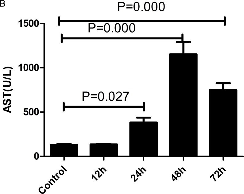
Results: During the progression of acute liver injury, the ALR levels increased slightly in early stage and significantly decreased in late stage in mice. Treatment with an ALR plasmid via tail vein injection protected mice against acute liver injury. The protective effect of ALR relied on the induction of autophagy, which was supported by the following evidence: (1) ALR overexpression directly induced autophagy flux in vitro and in vivo; and (2) ALR treatment suppressed apoptosis and promoted proliferation in mice exposed to CCl4, but the inhibition of autophagy reversed these effects. More importantly, the ALR levels decreased in patients with LC and ALF compared with normal controls.

Conclusion: We demonstrated that ALR ameliorated liver injury via an autophagic mechanism, which indicates a potential therapeutic application for liver injury.

Keywords: Pathology Section; apoptosis; augmenter of liver regeneration; autophagy; liver injury; proliferation.


Conflict of interest statement

CONFLICTS OF INTEREST

The authors declare no conflicts of interest.

Figures

Figure 1
Figure 1. ALR expression is significantly decreased during the late stage in mice with CCl4-induced acute liver injury
Mice received an intraperitoneal injection of a mixture of carbon tetrachloride (CCl4, 50%) and oil (50%) at a dose of 2 mL/kg body weight. The mice in the control group were injected with oil only. A. H&E staining of liver samples was performed from the control and 12, 24, 48, 72 h after the CCl4 injection. B. Serum AST and ALT levels were detected from the control and 12, 24, 48, 72 h after the CCl4 injection. C. Gene expression of ALR was measured by qRT-PCR in the livers of the control, 12, 24, 48, 72h after CCl4 injection. The average target gene/HPRT ratios for each experimental group were plotted. D. Protein expression of ALR was measured by western blot in the livers of the control and the 12, 24, 48, 72h after CCl4 injection. A representative blot from each group is shown. Densitometry analysis of the proteins was performed for each sample.
Figure 2
Figure 2. ALR protects mice against CCl4-induced acute liver injury
The ALR plasmid (10 mg/kg) or pcDNA3.0 (10 mg/kg) was injected into the tail vein 6 h before CCl4 exposure. The mice were sacrificed 48 h after the CCl4 injection, and control mice were injected with oil only. A. H&E staining of livers was performed in control mice, CCl4-treated mice and ALR/CCl4-treated mice.B. Serum AST and ALT levels were measured in control mice, CCl4-treated mice and ALR/CCl4-treated mice.
Figure 3
Figure 3. ALR promotes autophagy in CCl4-induced acute liver injury in vitro
The ALR plasmid (1 μg/ml) or pcDNA 3.0 (1 μg/ml) was transfected into AML12 cells. Cells were then cultured in RPMI-1640 for 6 h, followed by treatment with CCl4 (10 mmol/L) for 48 h. The control cells were treated with DMSO only. A. GFP-LC3B plasmids (1 μg/ml) were transfected into AML12 cells to observe the formation of autophagosomes. The green puncta indicated the presence of autophagosomes in control cells, CCl4-treated cells and ALR/CCl4-treated cells. B. The expression of LC3B, Atg5, p62 and ALR was measured by western blotting in control cells, CCl4-treated cells and ALR/CCl4-treated cells. A representative blot for two samples from each group is shown. Densitometry analysis of the proteins was performed for each sample.
Figure 4
Figure 4. ALR promotes autophagy in mice with CCl4-induced acute liver injury
The ALR plasmid (10 mg/kg) or pcDNA 3.0 (10 mg/kg) was injected into the tail vein 6 h before CCl4 exposure. The mice were sacrificed 48 h after CCl4 injection, and control mice were injected with oil only. The sections from control mice, CCl4-treated mice and ALR/CCl4-treated mice were observed in an inverted fluorescence microscope and an electron microscope. The expression of LC3B, Atg5, Atg7, Beclin-1, p62 and ALR was measured by western blotting in control mice, CCl4-treated mice and ALR/CCl4-treated mice. A representative blot for two samples from each group is shown. Densitometry analysis of the proteins was performed for each sample.
Figure 5
Figure 5. ALR suppresses apoptosis in mice with CCl4-induced acute liver injury through autophagic mechanisms
The ALR plasmid (10 mg/kg) or pcDNA 3.0 (10 mg/kg) was injected into the tail vein 6 h before CCl4 exposure. The suppression of autophagy was achieved with a tail vein injection of 3-methyladenine (3-MA, 1 mg/kg) 2 h before CCl4 exposure. The mice were sacrificed 48 h after CCl4 injection, and the control mice were injected with oil only. Apoptotic cells were stained in control mice, CCl4-treated mice, ALR/CCl4-treated mice, CCl4/3-MA-treated mice and ALR/CCl4/3-MA-treated mice with a TUNEL apoptosis detection kit (KeyGEN BioTECH, Nanjing, China). All of the images were obtained on an inverted fluorescence microscope.
Figure 6
Figure 6. ALR promotes proliferation in mice with CCl4-induced acute liver injury through autophagic mechanisms
The ALR plasmid (10 mg/kg) or pcDNA3.0 (10 mg/kg) was injected into tail vein 6 h before CCl4 exposure. Suppression of autophagy was achieved by tail vein injection of 3-methyladenine (3-MA, 1 mg/kg) 2 h before CCl4 exposure. The mice were sacrificed 48 h after CCl4 exposure, and the control mice were injected with oil only. The expression of proliferation-related proteins, including PCNA, CyclinA1, CyclinD1 and CyclinE1 was measured by western blotting in control mice, CCl4-treated mice, ALR/CCl4-treated mice, CCl4/3-MA-treated mice and ALR/CCl4/3-MA-treated mice. A representative blot for two samples from each group is shown. Densitometry analysis of the proteins was performed for each sample.
Figure 7
Figure 7. ALR expression is decreased in the liver of patients with LC and ALF
The expression of LC3B, Atg5, p62 and ALR was measured by western blotting in normal controls and patients with LC and ALF. A representative blot for two samples from each group is shown. Densitometry analysis of the proteins was performed for each sample.

References

    1. Khoury T, Rmeileh AA, Yosha L, Benson AA, Daher S, Mizrahi M. Drug Induced Liver Injury: Review with a Focus on Genetic Factors, Tissue Diagnosis, and Treatment Options. J Clin Transl Hepatol. 2015;3(2):99–108. - PMC - PubMed
    1. Oh IS, Park SH. Immune-mediated Liver Injury in Hepatitis B Virus Infection. Immune Netw. 2015;15(4):191–198. - PMC - PubMed
    1. Ren F, Zhang L, Zhang X, Shi H, Wen T, Bai L, Zheng S, Chen Y, Chen D, Li L, Duan Z. Inhibition of glycogen synthase kinase 3β promotes autophagy to protect mice from acute liver failure mediated by peroxisome proliferator-activated receptor α. Cell Death Dis. 2016;7:e2151. - PMC - PubMed
    1. Hagiya M, Francavilla A, Polimeno L. Cloning and sequence analysis of the rat augmenter of liver regeneration(ALR) gene: expression of biologically active recombinant ALR and demonstration of tissue distribution. Proc Natl Acad Sci USA. 1994;91(17):8142–8146. - PMC - PubMed
    1. Ilowski M, Putz C, Weiss TS, Brand S, Jauch KW, Hengstler JG, Thasler WE. Augmenter of liver regeneration causes different kinetics of ERK1/2 and Akt/PKB phosphorylation than EGF and induces hepatocyte proliferation in an EGF receptor independent and liver specific manner. Biochem Biophys Res Commun. 2010;394(4):915–920. - PubMed

MeSH terms

Substances