Review
. 2017 Jan 6:57:437-454.
doi: 10.1146/annurev-pharmtox-010716-105001.

Mitochondrial Mechanisms of Neuronal Cell Death: Potential Therapeutics

Affiliations
Review

Mitochondrial Mechanisms of Neuronal Cell Death: Potential Therapeutics

Ted M Dawson et al. Annu Rev Pharmacol Toxicol. .

Abstract

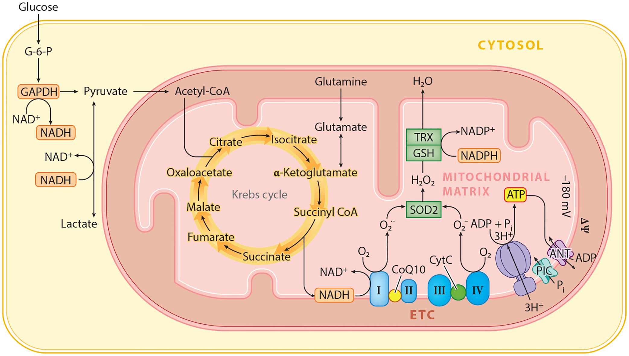
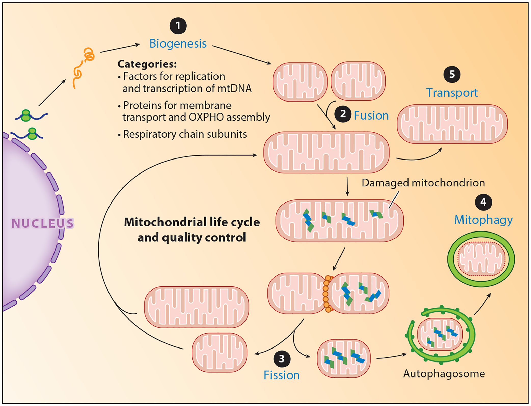
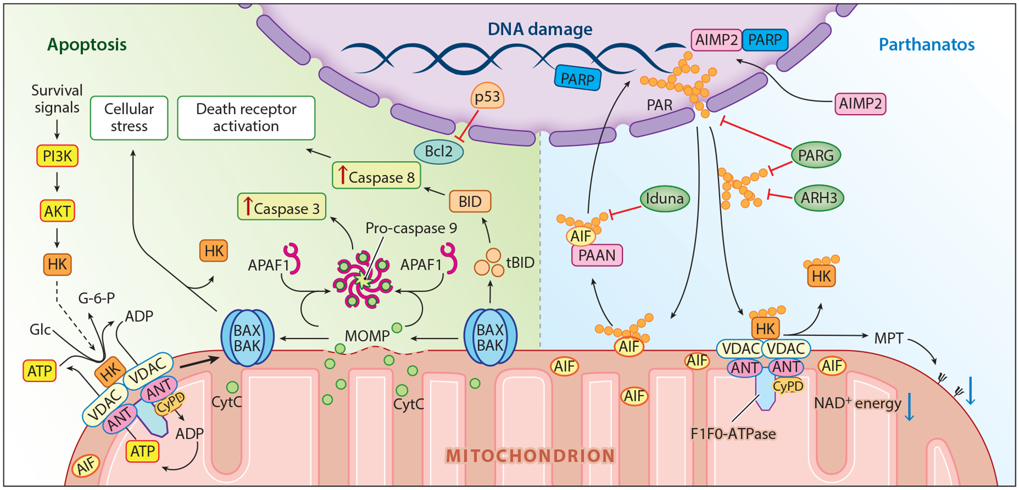
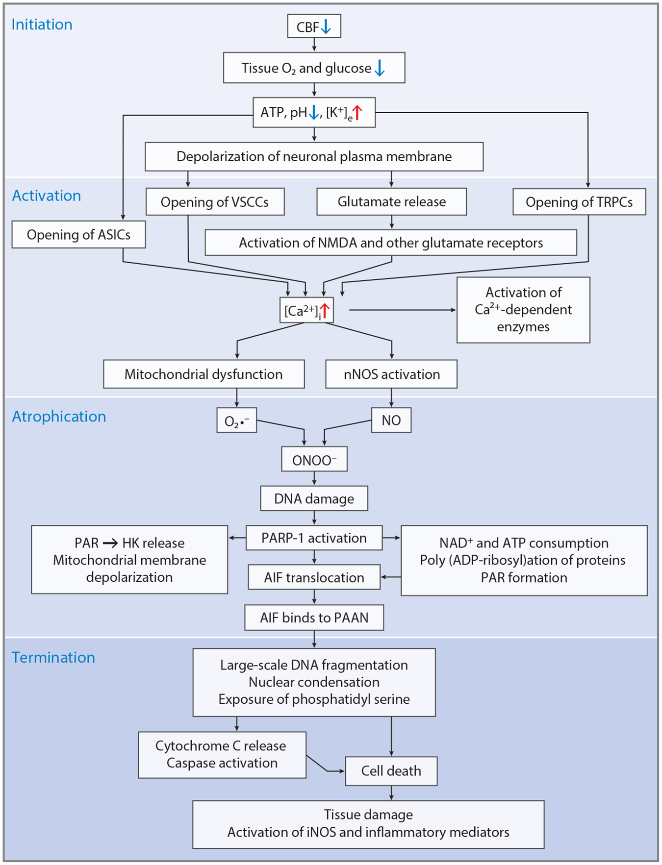
Mitochondria lie at the crossroads of neuronal survival and cell death. They play important roles in cellular bioenergetics, control intracellular Ca2+ homeostasis, and participate in key metabolic pathways. Mutations in genes involved in mitochondrial quality control cause a myriad of neurodegenerative diseases. Mitochondria have evolved strategies to kill cells when they are not able to continue their vital functions. This review provides an overview of the role of mitochondria in neurologic disease and the cell death pathways that are mediated through mitochondria, including their role in accidental cell death, the regulated cell death pathways of apoptosis and parthanatos, and programmed cell death. It details the current state of parthanatic cell death and discusses potential therapeutic strategies targeting initiators and effectors of mitochondrial-mediated cell death in neurologic disorders.

Keywords: apoptosis; apoptosis-inducing factor; neurodegeneration; parthanatos; poly (ADP-ribose) polymerase; stroke.


Conflict of interest statement

DISCLOSURE STATEMENT

T.M.D. and V.L.D. are founders of Valted, LLC and hold an ownership equity interest in the company. This arrangement has been reviewed and approved by the Johns Hopkins University in accordance with its conflict of interest policies.

Figures

Figure 1
Figure 1
Mitochondria play vital cellular functions including oxidative phosphorylation through the Krebs cycle and the electron transport chain. Abbreviations: ADP, adenosine diphosphate; ANT, adenine nucleotide transporter; ATP, adenosine triphosphate; CoQ10, coenzyme Q 10; CytC, cytochrome C; G-6-P, glucose-6-phosphate; ETC, electron transport chain; GSH, glutathione; H2O2, hydrogen peroxide; mV, millivolt; ΔΨ, mitochondrial membrane potential; NAD+, nicotinamide adenine dinucleotide; NADP+, nicotinamide adenine dinucleotide phosphate; O2, oxygen; Pi, phosphate; PIC, phosphate channel; O2•−, superoxide anion; SOD2, superoxide dismutase 2; TRX, thioredoxin; I, complex I; II, complex II; III, complex III; IV, complex IV.
Figure 2
Figure 2
The life cycle of mitochondria. Mitochondrial quality control is a tightly controlled, multistep process to meet the metabolic demands of the cell. ❶ Biogenesis coordinates the synthesis of new mitochondria. ❷ Fusion leads to joining of mitochondria ❸ whereas fission functions in part to segregate damaged mitochondria, which are engulfed by the autophagosome and degraded via ❹ mitophagy. In the nervous system, mitochondria are ❺ transported to the distal ends of axons and dendrites. Abbreviations: mtDNA, mitochondrial DNA; OXPHO, oxidative phosphorylation.
Figure 3
Figure 3
Mitochondrial-regulated cell death. Key players in apoptosis (left) and parthanatos (right) are highlighted. Abbreviations: AIF, apoptosis-inducing factor; AIMP2, aminoacyl-tRNA synthetase complex interacting multifunctional protein-2; AKT, protein kinase B; ANT, adenine nucleotide transporter; APAF1, apoptotic peptidase-activating factor 1; ARH3, ADP-ribosyl-acceptor hydrolase 3; ATP, adenosine triphosphate; BAK, Bcl-2 homologous antagonist/killer; BAX, Bcl-2-associated X; Bcl-2, B cell lymphoma 2; BID, BH3 (Bcl-2 homology 3) interacting-domain death agonist; CyPD, cyclophilin D; CytC, cytochrome C; Glc, glucose; G-6-P, glucose-6-phosphate; HK, hexokinase; Ψ, mitochondrial membrane potential; MOMP, mitochondrial outer membrane permeability; MPT, mitochondrial permeability transition; NAD+, nicotinamide adenine dinucleotide; PAAN, parthanatos AIF-associated nuclease; PAR, poly (ADP-ribose); PARG, PAR glycohydrolase; PARP, PAR polymerase; PI3K, phosphatidylinositol-3-kinase; p53, tumor suppressor p53; tBID, truncated BID.
Figure 4
Figure 4
Mechanisms of neuronal cell death during stroke. Neuronal cell death occurs in a four-step process involving initiation due to loss of cerebral blood flow; activation due to opening of a variety of calcium channels; atrophication, which involves the activation of calcium-dependent enzymes and oxidative and nitrosative stress, which leads to parthanatos; and, ultimately, death through large-scale DNA fragmentation and caspase activation. Tissue damage continues and is amplified through inflammatory mediators. Other causes of neuronal cell death may occur through similar mechanisms. Abbreviations: AIF, apoptosis-inducing factor; ASIC, acid-sensitive ion channel; ATP, adenosine triphosphate; [Ca2+]i, intracellular calcium; CBF, cerebral blood flow; DNA, deoxyribonucleic acid; HK, hexokinase; iNOS, inducible nitric oxide synthase; [K+]e, extracellular potassium; NAD+, nicotinamide adenine dinucleotide; NMDA, N-methyl-d-aspartate; NO, nitric oxide; nNOS, neuronal NO synthase; O2, oxygen; O2•−, superoxide anion; ONOO, peroxynitrite; PAAN, parthanatos AIF-associated nuclease; PAR, poly (ADP-ribose); PARP-1, PAR polymerase-1; TRPC, transient receptor potential channel; VSCC, voltage-sensitive cation channel.

References

    1. McGovern Inst. Brain Res. MIT. 2014. Brain Disorders: By the Numbers. Cambridge, MA: MIT. https://mcgovern.mit.edu/brain-disorders/by-the-numbers
    1. Di Carlo A 2009. Human and economic burden of stroke. Age Ageing 38:4–5 - PubMed
    1. Kowal SL, Dall TM, Chakrabarti R, Storm MV, Jain A. 2013. The current and projected economic burden of Parkinson’s disease in the United States. Mov. Disord 28:311–18 - PubMed
    1. Nath S, Villadsen J. 2015. Oxidative phosphorylation revisited. Biotechnol. Bioeng 112:429–37 - PubMed
    1. Paul VD, Lill R. 2015. Biogenesis of cytosolic and nuclear iron-sulfur proteins and their role in genome stability. Biochim. Biophys. Acta 1853:1528–39 - PubMed

Publication types