Review
. 2017 Jan 7;10(1):10.
doi: 10.1186/s13045-016-0373-z.

Natural compounds targeting major cell signaling pathways: a novel paradigm for osteosarcoma therapy

Affiliations
Review

Natural compounds targeting major cell signaling pathways: a novel paradigm for osteosarcoma therapy

Pablo Angulo et al. J Hematol Oncol. .

Abstract

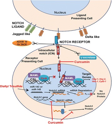
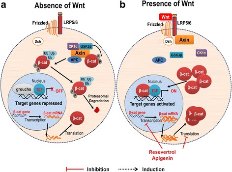
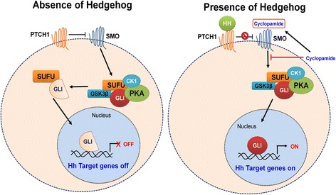
Osteosarcoma is the most common primary bone cancer affecting children and adolescents worldwide. Despite an incidence of three cases per million annually, it accounts for an inordinate amount of morbidity and mortality. While the use of chemotherapy (cisplatin, doxorubicin, and methotrexate) in the last century initially resulted in marginal improvement in survival over surgery alone, survival has not improved further in the past four decades. Patients with metastatic osteosarcoma have an especially poor prognosis, with only 30% overall survival. Hence, there is a substantial need for new therapies. The inability to control the metastatic progression of this localized cancer stems from a lack of complete knowledge of the biology of osteosarcoma. Consequently, there has been an aggressive undertaking of scientific investigation of various signaling pathways that could be instrumental in understanding the pathogenesis of osteosarcoma. Here, we review these cancer signaling pathways, including Notch, Wnt, Hedgehog, phosphatidylinositol-4,5-bisphosphate 3-kinase (PI3K)/AKT, and JAK/STAT, and their specific role in osteosarcoma. In addition, we highlight numerous natural compounds that have been documented to target these pathways effectively, including curcumin, diallyl trisulfide, resveratrol, apigenin, cyclopamine, and sulforaphane. We elucidate through references that these natural compounds can induce cancer signaling pathway manipulation and possibly facilitate new treatment modalities for osteosarcoma.

Keywords: Ezrin; Natural compounds; Osteosarcoma; Signaling pathways.


Figures

Fig. 1
Fig. 1
Chemical structure of the phytochemicals
Fig. 2
Fig. 2
Notch signaling pathway. Ligand from the presenting cell binds to the notch receptor on the receiving cell. Notch extracellular truncated (NEXT) domain is cleaved by ADAM metalloprotease and γ-secretase yielding the notch intracellular domain (NICD). NICD is translocated to the nucleus where it complexes with transcription factor CSL 9 CBF1/suppressor of hairless/Lag 1 and transcriptional coactivator of the mastermind-like proteins (MAML). The complex can then activate target gene transcription. Diallyl trisulfide (DATS) treatment increases expression of tumor suppressor microRNAs: miR-143 and miR-145. MicroRNAs bind to Notch1 mRNA and results in mRNA degradation with no translation of Notch1 protein. Curcumin downregulates transcription and translation Notch1 and downstream genes Hes-1, Hey-1, and Hey-2 of the nucleus
Fig. 3
Fig. 3
Wnt signaling pathway. a In the absence of the Wnt glycoprotein, β-catenin is degraded after being ubiquitinated and phosphorylated by the destruction complex. Target genes in the nucleus are not activated. b In the presence of Wnt, the glycoprotein binds to the extracellular transmembrane Frizzled receptor family (Fz and LRP5/6). Thereafter, the signal activates the protein Dishevelled (Dsh/DV1) in the cytoplasm. This binding results in disrupting the β-catenin destruction complex of various proteins including: axin, casein kinase 1α, adenomatous polyposis coli (APC), protein phosphatase 2A (PP2A), and glycogen synthase kinase 3 (GSK–3β). β-catenin translocates to nucleus where it can act as transcriptional coactivator of transcription factors of TCF/LEF family. Resveratrol and apigenin decrease protein expression of β-catenin
Fig. 4
Fig. 4
Hedgehog signaling pathway. a In the absence of Hh ligand, PTCH prevents activation of SMO. SMO cannot inhibit protein kinases including PKA, GSK-3β, and CK1. These protein kinases phosphorylate GLI protein in complex with SUFU resulting in cleavage of GLI into a repressed form. The repressed form will translocate to the nucleus inhibiting Hh target gene expression. b Hh ligand binds PTCH1 (transmembrane receptor). Smoothened (SMO) is relieved and inhibits proteolytic cleavage of GLI protein resulting in an active form. The active GLI protein translocates to nucleus and activates transcription factors. Cyclopamine binds to SMO preventing signal transduction to GLIS
Fig. 5
Fig. 5
PI3K-AKT-mTOR and RAS-RAF-MEK-ERK pathways. Growth factor binds to epidermal growth factor receptor and can proceed by two different pathways. For the PI3 pathway, the ligand activates tyrosine kinase receptor activity resulting in phosphorylation of receptor. PI3K binds to phosphorylated receptor and becomes activated. PI3K then binds to PIP2 on inner membrane and phosphorylates PIP2 to PIP3. PIP3 activates AKT via PDK1. AKT can then phosphorylate and activate protein mTOR which results in cell growth, cell proliferation, and cell survival. For the RAS-RAF pathway, growth factor binding to tyrosine kinase receptor activates RAS which in turn activates RAF. RAF activates MEK which phosphorylates ERK to decrease apoptosis and increase cell proliferation and growth. The compound sulforaphane suppresses the phosphorylation of AKT and ERK
Fig. 6
Fig. 6
Ezrin pathway. While in the dormant form, ezrin with N-terminal ezrin/radixin/moesin (ERM)-associated domain (N-ERMAD) associates in the cytoplasm with carboxy-ERMAD (C-ERMAD). Ezrin then becomes phosphorylated at various sites resulting in transformation to the active form. The C-terminal of transmembrane proteins as well as C-ERMAD binds with the N-terminal of activated ezrin. In addition, ezrin can serve as a linker protein between specific membranous proteins and F-actin via ERM-binding phosphoprotein 50 (EBP50). Guanosine diphosphate inhibitor (GDI) from the Rho-GDI complex is displaced by activated ezrin. This displacement can then stimulate PI4P5 kinase activity which is catalyzed by GDP/GTP exchange factor (GEF). Thereafter, PI4P5 kinase can act on PIP to convert PIP to phosphatidylinositol (4,5)-bisphosphate (PIP2). Thus, PIP2 sequentially converts dormant ezrin into the active form
Fig. 7
Fig. 7
Jak/STAT pathway. The corresponding cytokine or growth factor ligand binds to the cell surface receptor prompting activated JAK2 protein to phosphorylate tyrosine residues in the cytoplasmic domain of the receptor. More so, JAK2 also phosphorylates recruited signal transducer and activator of transcription (STAT) which results in STAT dimerization via conserved Src homology 2 (SH2) domains. STAT dimers then translocate to the nucleus where they induce transcription of target genes. In addition, JAK acts as a docking site for SH2 containing adapter proteins including Src homology 2 domain-containing phosphatase 2 (SHP2), growth factor receptor bound protein-2 (GRB2), and Src homology 2 domain-containing transforming protein (SHC). GRB2 which is associated with Son of Sevenless (SOS) can bind the tyrosine phosphorylated receptor directly or indirectly by way of the Src homology 2 domain-containing protein (SHC). This binding results in the translocation of SOS to the membrane. At the membrane, SOS exchanges GDP for GTP on Ras guanine nucleotide-binding proteins. Ras-GTP can then activate MAPK cascade. Aside from RAS, JAK/STAT also interacts with PI3 and AKT pathways [65]. Under normal conditions gene expression is regulated by negative feedback mechanisms including the production of the negative regulator suppressors of cytokine signaling (SOCS)

References

    1. Messerschmitt PJ, Garcia RM, Abdul-Karim FW, Greenfield EM, Getty PJ. Osteosarcoma. J Am Acad Orthop Surg. 2009;17:515–527. doi: 10.5435/00124635-200908000-00005. - DOI - PubMed
    1. Klein MJ, Siegal GP. Osteosarcoma: anatomic and histologic variants. Am J Clin Pathol. 2006;125:555–581. doi: 10.1309/UC6KQHLD9LV2KENN. - DOI - PubMed
    1. Picci P. Osteosarcoma (osteogenic sarcoma) Orphanet J Rare Dis. 2007;2:6. doi: 10.1186/1750-1172-2-6. - DOI - PMC - PubMed
    1. Botter SM, Neri D, Fuchs B. Recent advances in osteosarcoma. Curr Opin Pharmacol. 2014;16:15–23. doi: 10.1016/j.coph.2014.02.002. - DOI - PubMed
    1. Jaffe N, Puri A, Gelderblom H. Osteosarcoma: evolution of treatment paradigms. Sarcoma. 2013;2013:203531. doi: 10.1155/2013/203531. - DOI - PMC - PubMed

Publication types

Substances

LinkOut - more resources