. 2017 Feb 17;292(7):2916-2923.
doi: 10.1074/jbc.M116.753574. Epub 2017 Jan 5.

Cardiolipin Regulates Mitophagy through the Protein Kinase C Pathway

Affiliations

Cardiolipin Regulates Mitophagy through the Protein Kinase C Pathway

Zheni Shen et al. J Biol Chem. .

Abstract

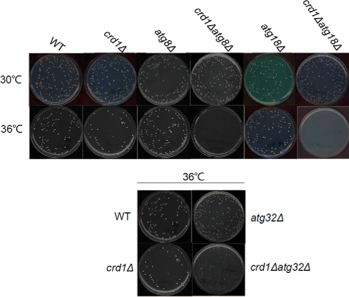
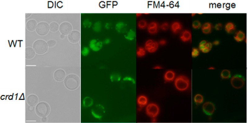
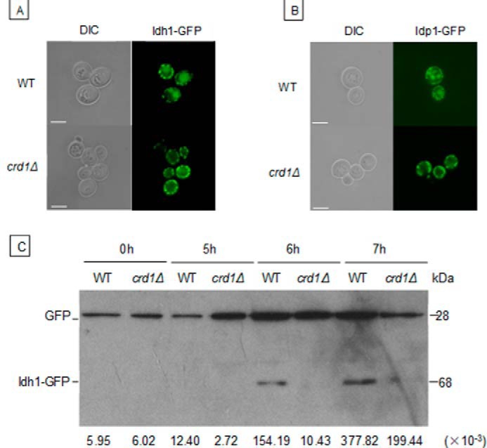
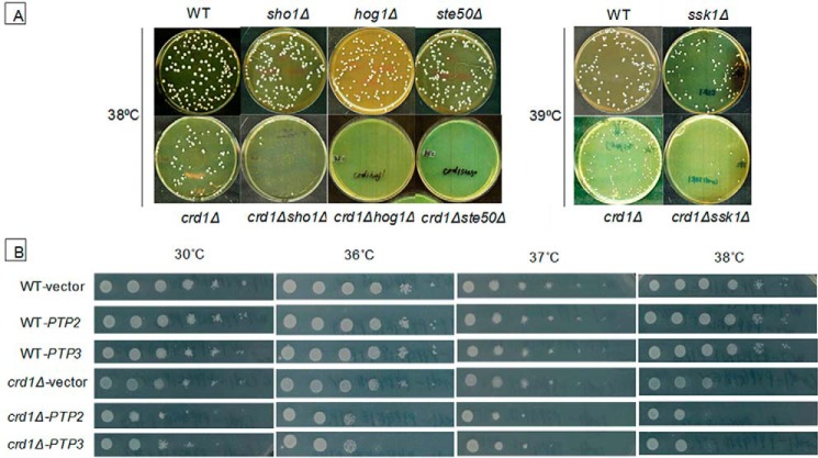
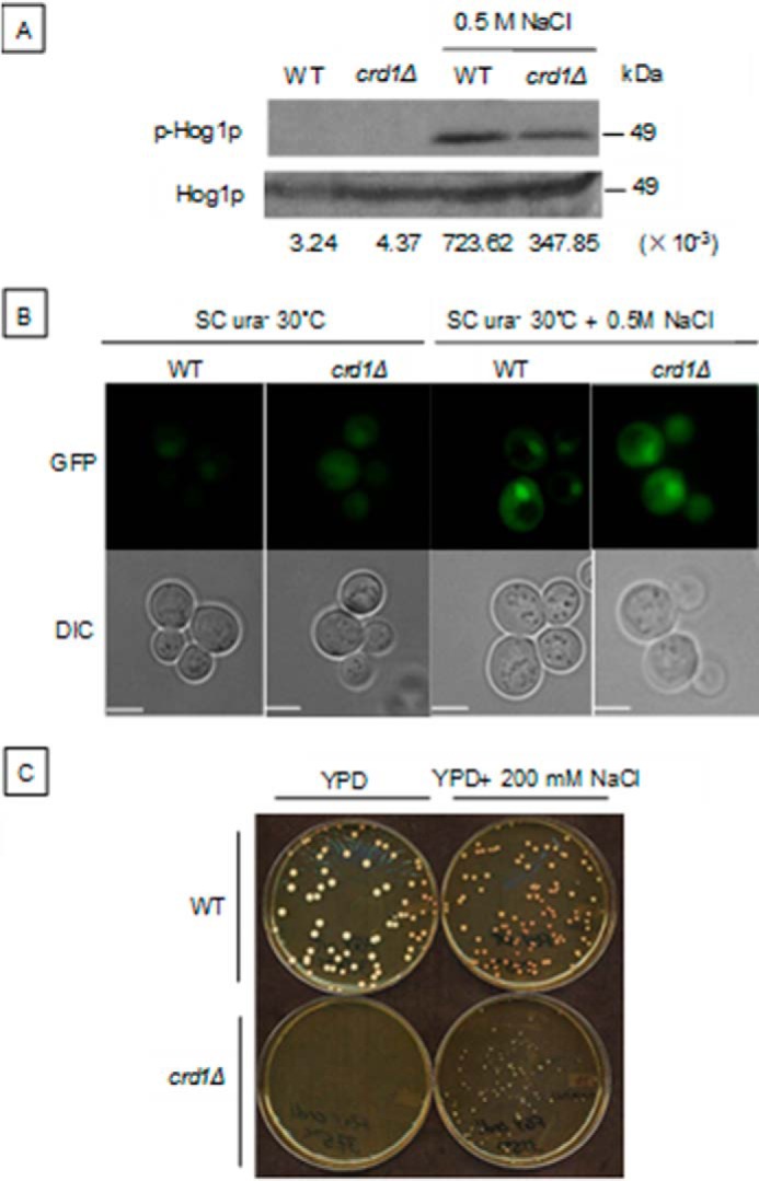
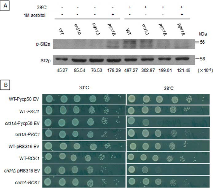
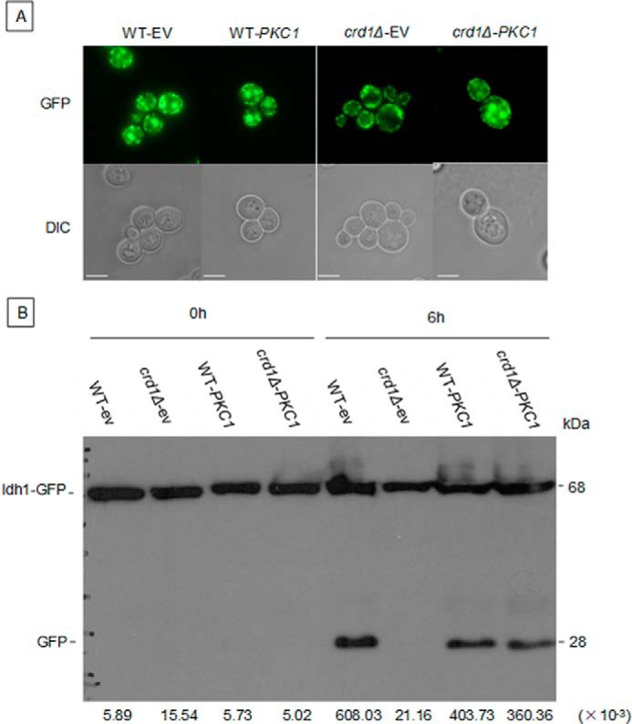
Cardiolipin (CL), the signature phospholipid of mitochondrial membranes, is important for cardiovascular health, and perturbation of CL metabolism is implicated in cardiovascular disease. Although the role of CL in mitochondrial function, biogenesis, and genome stability has been studied, recent findings indicate that it is essential for functions apart from mitochondrial bioenergetics. In this study, we report that mitophagy is perturbed in CL-deficient yeast cells. Mutants of autophagy/mitophagy genes ATG8, ATG18, and ATG32 synthetically interact with CL synthase mutant crd1Δ. CL-deficient cells exhibited decreased GFP-tagged mitochondrial proteins inside the vacuole and decreased free GFP, consistent with decreased mitophagy. Both PKC and high osmolarity glycerol (HOG) MAPK pathways were shown previously to be required for mitophagy. Activation of both MAPKs was defective in CL-deficient cells. Deletion of HOG pathway genes SHO1, SSK1, STE50, and HOG1 exacerbated crd1Δ growth. 1 m sorbitol and 0.2 m NaCl, which induce the HOG pathway, rescued growth of the mutant. Activation of the MAPK Slt2p was defective in crd1Δ cells, and up-regulation of the PKC pathway by expression of the PKC1R398P gene, which encodes constitutively activated Pkc1p, rescued crd1Δ growth and mitophagy defects. These findings indicate that loss of CL impairs MAPK pathway activation, and decreased activation of the PKC pathway leads to defective mitophagy.

Keywords: cardiolipin; mitochondria; mitogen-activated protein kinase (MAPK); mitophagy; protein kinase C (PKC).


Conflict of interest statement

The authors declare that they have no conflicts of interest with the contents of this article

Figures

FIGURE 1.
FIGURE 1.
Deletion of autophagy/mitophagy genes exacerbates crd1Δ growth defect at elevated temperature. Cells in the FGY genetic background were precultured in YPD at 30 °C to the mid-log phase. About 200 cells of each strain were plated on YPD and incubated at 30 °C or 36 °C for 3 days.
FIGURE 2.
FIGURE 2.
Delivery of mitochondria to the vacuole is decreased in crd1Δ cells. FGY WT and crd1Δ cells expressing a genomic copy of GFP-tagged IDH1 were cultured in YPD medium to the mid-log phase at 30 °C and then shifted to 39 °C for 8 h and observed under fluorescence microscopy. All images were taken at the same magnification (×1000). Bar, 2 μm.
FIGURE 3.
FIGURE 3.
Decreased mitophagy in crd1Δ cells. WT and crd1Δ cells in the BY4742 genetic background expressing an endogenous Idh1-GFP (A) or exogenous Idp1-GFP (B) were cultured in SC medium to the mid-log phase at 30 °C and shifted to SL for 18 h. Cells were observed under fluorescence microscopy. C, WT and crd1Δ cells carrying the GFP-tagged IDH1 gene were precultured in SC medium to the mid-log phase at 30 °C and shifted to SL medium for the indicated time. Aliquots were analyzed by immunoblotting with anti-YFP antibody, and the positions of full-length Idh1-GFP and free GFP are indicated. Images in A and B were taken at the same magnification (×1000). Bar, 2 μm. Numbers at the bottom of C show the density ratios for GFP to Idh1-GFP in each lane. DIC, differential interference contrast.
FIGURE 4.
FIGURE 4.
Down-regulation of the HOG pathway exacerbates the crd1Δ growth defect. A, cells in the BY4742 genetic background were precultured in YPD at 30 °C to the mid-log phase. 200 cells of each strain were plated on YPD plates and incubated at the indicated temperature for 3 days. B, cells were precultured in SC leu at 30 °C to the mid-log phase. Serial dilutions were spotted on SC leu plates (with the most diluted spot containing 20 cells), and the plates were incubated at the indicated temperature for 2 days.
FIGURE 5.
FIGURE 5.
Decreased Hog1p phosphorylation in the crd1Δ mutant. A, cells in the BY4742 genetic background were precultured in YPD at 30 °C to the mid-log phase and treated with 0.5 m NaCl for 5 min. Dual phosphorylated (activated) and total Hog1 proteins were detected by Western blotting, as described previously (8). B, WT and crd1Δ cells expressing Hog1-GFP on the plasmid pPS1739 were grown in SC ura to an A550 of 1, treated with 0.5 m NaCl for 5 min, and then observed using fluorescence microscopy. C, WT and crd1Δ cells in the FGY background were precultured in YPD liquid at 30 °C to the mid-log phase. 200 cells were plated on a YPD plate with or without 200 mm NaCl. Plates were then incubated at 37.5 °C for 3 days. Numbers at the bottom of A show the density ratios for p-Hog1p to Hog1p in each lane. Images in B were taken at the same magnification (×1000). Bar, 2 μm. DIC, differential interference contrast.
FIGURE 6.
FIGURE 6.
Up-regulation of the PKC pathway rescues the crd1Δ growth defect. A, cells in the BY4742 genetic background were precultured in liquid YPD at 30 °C to the mid-log phase, diluted to an A550 of 0.3, and treated with increased temperature or 1 m sorbitol for 2 h. Cell extracts were prepared and activated (dual phosphorylated), and total Slt2 protein was detected by Western blotting. B, cells expressing BCK1-20 and PKC1R398P plasmids were cultured in SC ura medium at 30 °C to the mid-log phase, diluted, spotted on SC ura plates, and incubated at 30 or 38 °C for 2 days. Numbers at the bottom of A show the density ratios for p-Slt2p to Slt2p in each lane.
FIGURE 7.
FIGURE 7.
Up-regulation of the PKC pathway restores mitophagy in crd1Δ. BY WT-IDH1-GFP and crd1-IDH1-GFP cells containing either an empty vector or a vector expressing PKC1R398P were cultured in SC ura medium to the mid-log phase at 30 °C, washed, and shifted to SL (to induce mitophagy). Aliquots were observed 18 h after shift under fluorescence microscopy (A), and full-length Idh1-GFP and free GFP were identified by Western blotting using anti-GFP antibody 6 h after shift (B). Images in A were taken at the same magnification (×1000). Bar, 2 μm. Numbers at the bottom of B show the density ratios for GFP to Idh1-GFP in each lane. DIC, differential interference contrast.

References

    1. Shen Z., Ye C., McCain K., and Greenberg M. L. (2015) The role of cardiolipin in cardiovascular health. Biomed Res. Int. 2015, 891707. - PMC - PubMed
    1. Barth P. G., Scholte H. R., Berden J. A., Van der Klei-Van Moorsel J. M., Luyt-Houwen I. E., Van 't Veer-Korthof E. T., Van der Harten J. J., and Sobotka-Plojhar M. A. (1983) An X-linked mitochondrial disease affecting cardiac muscle, skeletal muscle and neutrophil leucocytes. J. Neurol. Sci. 62, 327–355 - PubMed
    1. Bione S., D'Adamo P., Maestrini E., Gedeon A. K., Bolhuis P. A., and Toniolo D. (1996) A novel X-linked gene, G4.5. is responsible for Barth syndrome. Nat. Genet. 12, 385–389 - PubMed
    1. Valianpour F., Wanders R. J. A., Overmars H., Vreken P., Van Gennip A. H., Baas F., Plecko B., Santer R., Becker K., and Barth P. G. (2002) Cardiolipin deficiency in X-linked cardioskeletal myopathy and neutropenia (Barth syndrome, mim 302060): a study in cultured skin fibroblasts. J. Pediatrics 141, 729–733 - PubMed
    1. Schlame M., Towbin J. A., Heerdt P. M., Jehle R., DiMauro S., and Blanck T. J. J. (2002) Deficiency of tetralinoleoyl-cardiolipin in Barth syndrome. Ann. Neurol. 51, 634–637 - PubMed