. 2017 Mar 1;30(3):197-204.
doi: 10.1093/protein/gzw078.

Dual chemistry catalyzed by human acireductone dioxygenase

Affiliations

Dual chemistry catalyzed by human acireductone dioxygenase

Aditi R Deshpande et al. Protein Eng Des Sel. .

Abstract

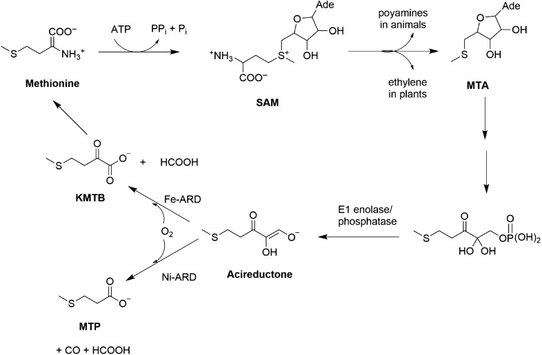
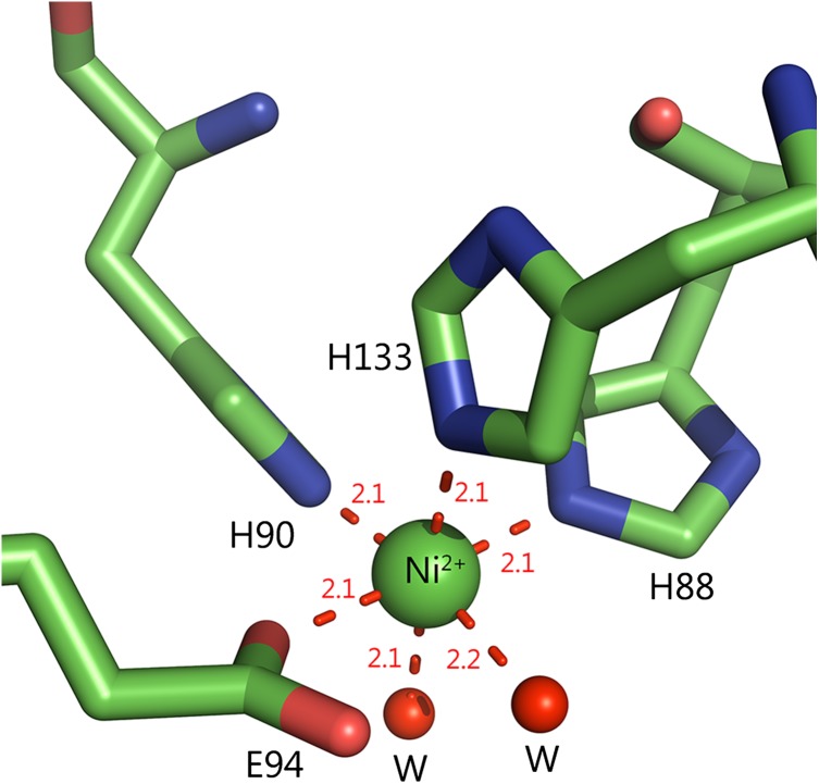
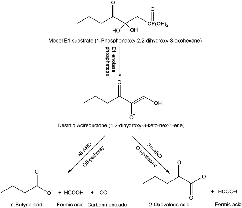
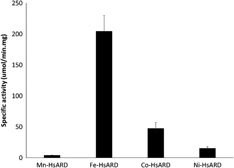
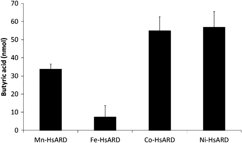
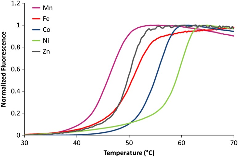
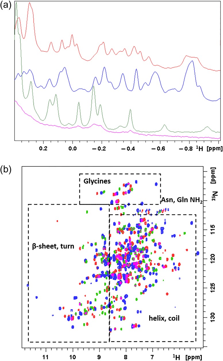
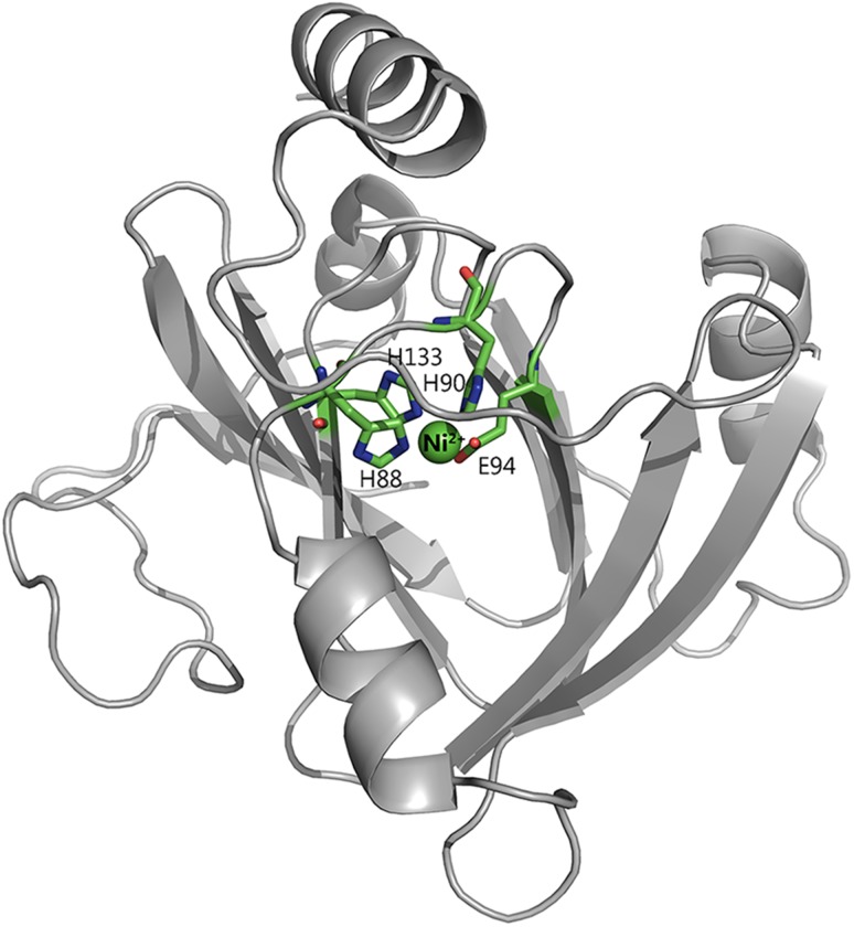
Acireductone dioxygenase (ARD) from the methionine salvage pathway of Klebsiella oxytoca is the only known naturally occurring metalloenzyme that catalyzes different reactions in vivo based solely on the identity of the divalent transition metal ion (Fe2+ or Ni2+) bound in the active site. The iron-containing isozyme catalyzes the cleavage of substrate 1,2-dihydroxy-3-keto-5-(thiomethyl)pent-1-ene (acireductone) by O2 to formate and the ketoacid precursor of methionine, whereas the nickel-containing isozyme uses the same substrates to catalyze an off-pathway shunt to form methylthiopropionate, carbon monoxide and formate. This dual chemistry was recently demonstrated in vitro by ARD from Mus musculus (MmARD), providing the first example of a mammalian ARD exhibiting metal-dependent catalysis. We now show that human ARD (HsARD) is also capable of metal-dependent dual chemistry. Recombinant HsARD was expressed and purified to obtain a homogeneous enzyme with a single transition metal ion bound. As with MmARD, the Fe2+-bound HsARD shows the highest activity and catalyzes on-pathway chemistry, whereas Ni2+, Co2+ or Mn2+ forms catalyze off-pathway chemistry. The thermal stability of the HsARD isozymes is a function of the metal ion identity, with Ni2+-bound HsARD being the most stable followed by Co2+ and Fe2+, and Mn2+-bound HsARD being the least stable. As with the bacterial ARD, solution NMR data suggest that HsARD isozymes can have significant structural differences depending upon the metal ion bound.

Keywords: Klebsiella oxytoca; acireductone; dual chemistry; mammalian; metal.


Figures

Fig. 1
Fig. 1
The MSP in Klebsiella oxytoca.
Fig. 2
Fig. 2
Active site metal coordination geometry. The active site of MmARD is shown with the Ni atom indicated as a green sphere and its protein ligands H88, H90, H133, E94 represented as sticks and two water molecules denoted as ‘W’ shown as red spheres forming a distorted octahedral coordination geometry. The metal coordination distances are shown as red dotted lines with distances measured in Å.
Fig. 3
Fig. 3
Sequence alignment of ARD homologs. The metal-binding residues are marked by arrows and the diamond represents the first methionine residue of N-terminal truncated HsARD denoted as SiPL which is implicated in replication of hepatitis C virus in non-permissive cell lines. Sequence alignment was performed using ClustalOmega.
Fig. 4
Fig. 4
ARD reactions using desthio E1 substrate to produce desthio-acireductone and the desthio on-pathway and off-pathway ARD chemistry products.
Fig. 5
Fig. 5
Specific activity of HsARD proteins bound to different metal ions. The assay was performed in argon-saturated 50 mM HEPES pH 7.0, 1 mM MgCl2 buffer with 0.6 µM E1 enzyme, 500 µM E1 substrate in a total volume of 1 mL. A controlled amount of ARD was added and the depletion of acireductone was monitored spectrophotometrically at 308 nm. The errors are represented as standard deviations of three replicates.
Fig. 6
Fig. 6
Off-pathway product analysis of different metal ion-bound HsARDs. The enzymatic reaction mixture contained argon-saturated 50 mM HEPES pH 7.0, 1 mM MgCl2 buffer with 0.3 µM E1 enzyme, 250 µM E1 substrate in a total volume of 1 mL. About 100 µL of this reaction mixture was loaded onto the organic acid column. The quantitation is based on a 1 mL reaction mixture. The errors are represented as standard deviations of three replicates.
Fig. 7
Fig. 7
Thermal stability of HsARD as a function of the bound metal ion. The samples for analysis contained 20 µM protein with 10× SYPRO Orange (Invitrogen) in final volume of 25 µL. The samples were heated at a ramp rate of 0.3°C/min and a temperature range of 25–90°C.
Fig. 8
Fig. 8
Solution NMR data of Mn2+, Fe2+, Co2+ and Ni2+-bound HsARD isozymes. (a) Upfield region of 800 MHz 1H NMR spectra of Mn-HsARD (pink), Fe-HsARD (red), Co-HsARD (blue) and Ni-HsARD (green) showing methyl resonances of amino acid side chains ring current-shifted by nearby aromatic residues. The different spectral patterns indicate differences in side chain packing in hydrophobic cores. (b) Overlay of the 2D 1H, 15N HSQC spectra of Mn-HsARD (pink), Co-HsARD (blue), Fe-HsARD (red) and Ni-HsARD (green). Different shift patterns indicate differences local hydrogen bonding and structure. All spectra were obtained at 25°C, pH 7.0 at 800 MHz 1H observe frequency.
Fig. 9
Fig. 9
Homology model of HsARD. Model is based on the crystal structure of Ni-MmARD (PDB ID: 5I8S). The Ni atom is indicated as a green sphere and the protein ligands H88, H90, H133, E94 are represented as sticks. The remaining two metal coordination sites are shown empty. In the case of MmARD, these are occupied by water molecules as indicated in Fig. 2.

References

    1. Chai S.C., Ju T., Dang M., Goldsmith R.B., Maroney M.J. and Pochapsky T.C. (2008) Biochemistry, 47, 2428–2438. - PMC - PubMed
    1. Chou H.Y., Lin Y.H., Shiu G.L., Tang H.Y., Cheng M.L., Shiao M.S. and Pai L.M. (2014) J. Biomed. Sci., 21, 64. - PMC - PubMed
    1. Crans D.C., Yang L., Gaidamauskas E., Kahn R., Jin W. , Simonis U. (2003) In Paramagnetic Resonance of Metallobiomolecules (Telser, J., Ed.), American Chemical Society, Washington D.C.
    1. Dai Y., Pochapsky T.C. and Abeles R.H. (2001) Biochemistry, 40, 6379–6387. - PubMed
    1. Dai Y., Wensink P.C. and Abeles R.H. (1999) J. Biol. Chem., 274, 1193–1195. - PubMed

Publication types