. 2017 Jan 24;114(4):764-769.
doi: 10.1073/pnas.1608839114. Epub 2017 Jan 6.

Activating mutations and translocations in the guanine exchange factor VAV1 in peripheral T-cell lymphomas

Affiliations

Activating mutations and translocations in the guanine exchange factor VAV1 in peripheral T-cell lymphomas

Francesco Abate et al. Proc Natl Acad Sci U S A. .

Abstract

Peripheral T-cell lymphomas (PTCLs) are a heterogeneous group of non-Hodgkin lymphomas frequently associated with poor prognosis and for which genetic mechanisms of transformation remain incompletely understood. Using RNA sequencing and targeted sequencing, here we identify a recurrent in-frame deletion (VAV1 Δ778-786) generated by a focal deletion-driven alternative splicing mechanism as well as novel VAV1 gene fusions (VAV1-THAP4, VAV1-MYO1F, and VAV1-S100A7) in PTCL. Mechanistically these genetic lesions result in increased activation of VAV1 catalytic-dependent (MAPK, JNK) and non-catalytic-dependent (nuclear factor of activated T cells, NFAT) VAV1 effector pathways. These results support a driver oncogenic role for VAV1 signaling in the pathogenesis of PTCL.

Keywords: VAV1; gene fusion; mutation; peripheral T-cell lymphoma.


Conflict of interest statement

The authors declare no conflict of interest.

Figures

Fig. 1.
Fig. 1.
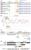
VAV1 fusion genes in PTCL. (A) Schematic representation of the domain structure of the VAV1 protein. (B) Schematic representation of the domain structures of the VAV1-S100A7, VAV1-THAP4, and VAV1-MYO1F fusion proteins. Ac, acidic domain; C1, C1 domain; recognition motif for diacylglycerol and phorbol esters, atypical; CH, calponin homology domain; DH, DBL homology; EF, pseudo-EF hand domain; nitrobindin, nitrobindin domain; PH, pleckstrin homology domain; SH2, Src homology 2 domain; SH3, Src homology 3 domain.
Fig. 2.
Fig. 2.
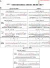
Recurrent VAV1 Δ778–786 mutation in PTCL. (A) Schematic representation of the domain structure of the VAV1 protein indicating the location of the VAV1 p.778delVGSTKYFGT (VAV1 Δ778–786) mutation. Each red circle is indicative of a PTCL mutant sample. (B) Genomic DNA and cDNA sequences corresponding to the intron 25–exon 26 genomic DNA and exon 25–exon 26 cDNA boundaries, respectively, in PTCL samples harboring the VAV1 p.778delVGSTKYFGT (VAV1 Δ778–786) mutation.
Fig. 3.
Fig. 3.
VAV1 intron 25–exon 26 deletion–induced missplicing and VAV1 Δ778–786 expression. (A) Genomic DNA sequences for PTCLs with verified intron 25–exon 26 indel mutations. Deleted genomic DNA sequences are indicated with orange dotted lines. Inserted nucleotides are indicated in red. Intron 25 nucleotides are shown in lowercase letters. Exon 26 nucleotides are indicated in capital letters. (B) VAV1 exon 26 splicing sequencer analysis. ESE, exonic splicing enhancer; ESS, exonic splicing silencer. P scores indicate the Z value for sequence over/underrepresentation in internal noncoding exons vs. pseudo exons. I scores indicate the Z value for sequence over/underrepresentation in internal noncoding exons vs. 5′ UTRs of intronless genes. Underrepresented octamers are assigned negative Z scores. Nine nucleotides corresponding to two overlapping octamers, GTATTTAT and TATTTATG, with strong exonic splicing silencer scores are boxed in orange. (C) Scheme of genomic DNA of wild-type VAV1 intron 25–exon 26 boundary indicating the intron 25 polypyrimidine tract and splice acceptor sequence (green box), exon 26 exonic splicing silencer (orange box), and the cryptic alternative splice acceptor site activated in PTCL samples harboring VAV1 intron 25–exon 26 indel mutations (blue box). Partial sequences of exons 25 and 26 (capital letters) and intron 25 (lowercase letters) are shown. (D) Schematic representation of a representative VAV1 intron 25–exon 26 mutation (g.81275_81301del) and the consequent missplicing-induced deletion (r.2473_2499del) and protein product (p.Val778_Thr786del; VAV1 Δ835–845).
Fig. 4.
Fig. 4.
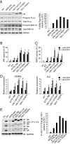
VAV1 fusions and the VAV1 Δ778–786 mutation induce increased VAV1 signaling in T cells. (A) Analysis of ERK1/2 and PLCγ1 phosphorylation in J.VAV1 cells upon expression of VAV1 fusion proteins or the VAV1 Δ778–786 intragenic deletion mutant. The VAV1 Δ835–845 C-terminal SH3 domain deletion mutant is used as positive control for VAV1 activity. (B and C) Reporter assays analyzing JNK (B) and NFAT activity (C) in JURKAT cells expressing wild-type, mutant, or fusion VAV1 proteins in basal conditions and upon stimulation with anti-CD3. A.U., arbitrary units. (D) Quantitative RT-PCR analysis of CD40L and IL2 NFAT target genes in JURKAT cells expressing wild-type, mutant, or fusion VAV1 proteins in basal conditions and upon stimulation with anti-CD3. (E) Immunoprecipitation/Western blot analysis of VAV1 Tyr174 phosphorylation in J.VAV1 cells upon expression of wild-type and mutant or fusion VAV1 proteins shows increased activated phosphorylation of VAV1 mutants and fusion proteins compared with wild-type control. Bar graphs in BD show mean values, and error bars represent the SD. Values are indicative of results in triplicate samples in a representative example of three independent experiments. P values were calculated using two-tailed Student’s t test; *P < 0.05 relative to nonstimulated wild-type VAV1-expressing cells; #P < 0.05 relative to stimulated wild-type VAV1-expressing cells. EV, empty vector; IB, immunoblotting; IP, immunoprecipitation.

References

    1. de Leval L, Gaulard P. Pathology and biology of peripheral T-cell lymphomas. Histopathology. 2011;58(1):49–68. - PubMed
    1. Rüdiger T, Müller-Hermelink HK. [WHO-classification of malignant lymphomas] Radiologe. 2002;42(12):936–942. German. - PubMed
    1. Armitage JO. The aggressive peripheral T-cell lymphomas: 2015. Am J Hematol. 2015;90(7):665–673. - PubMed
    1. Savage KJ, Ferreri AJ, Zinzani PL, Pileri SA. Peripheral T-cell lymphoma—Not otherwise specified. Crit Rev Oncol Hematol. 2011;79(3):321–329. - PubMed
    1. Federico M, et al. Clinicopathologic characteristics of angioimmunoblastic T-cell lymphoma: Analysis of the International Peripheral T-Cell Lymphoma Project. J Clin Oncol. 2013;31(2):240–246. - PMC - PubMed

Publication types