. 2017 Jan;16(1):76-87.
doi: 10.1158/1535-7163.MCT-16-0205. Epub 2016 Nov 15.

Novel p21-Activated Kinase 4 (PAK4) Allosteric Modulators Overcome Drug Resistance and Stemness in Pancreatic Ductal Adenocarcinoma

Affiliations

Novel p21-Activated Kinase 4 (PAK4) Allosteric Modulators Overcome Drug Resistance and Stemness in Pancreatic Ductal Adenocarcinoma

Amro Aboukameel et al. Mol Cancer Ther. 2017 Jan.

Abstract

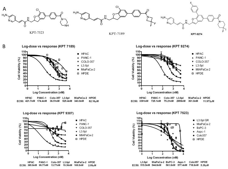
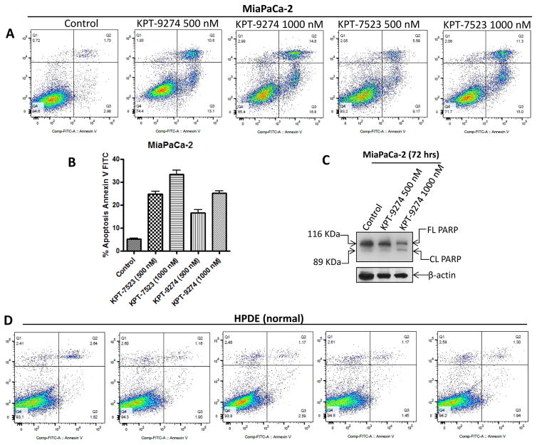
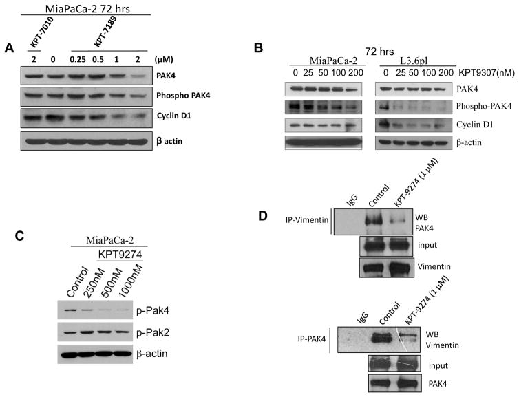
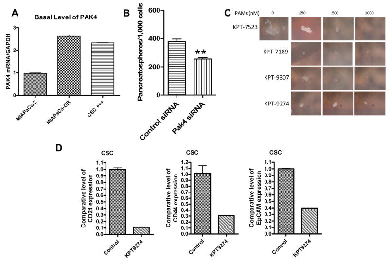
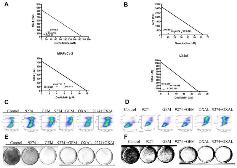
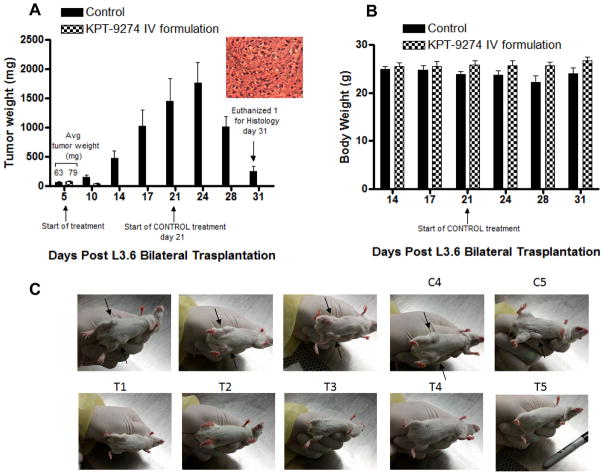
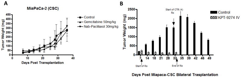
The p21-activated kinase 4 (PAK4) is a key downstream effector of the Rho family GTPases and is found to be overexpressed in pancreatic ductal adenocarcinoma (PDAC) cells but not in normal human pancreatic ductal epithelia (HPDE). Gene copy number amplification studies in PDAC patient cohorts confirmed PAK4 amplification making it an attractive therapeutic target in PDAC. We investigated the antitumor activity of novel PAK4 allosteric modulators (PAM) on a panel of PDAC cell lines and chemotherapy-resistant flow-sorted PDAC cancer stem cells (CSC). The toxicity and efficacy of PAMs were evaluated in multiple subcutaneous mouse models of PDAC. PAMs (KPT-7523, KPT-7189, KPT-8752, KPT-9307, and KPT-9274) show antiproliferative activity in vitro against different PDAC cell lines while sparing normal HPDE. Cell growth inhibition was concurrent with apoptosis induction and suppression of colony formation in PDAC. PAMs inhibited proliferation and antiapoptotic signals downstream of PAK4. Co-immunoprecipitation experiments showed disruption of PAK4 complexes containing vimentin. PAMs disrupted CSC spheroid formation through suppression of PAK4. Moreover, PAMs synergize with gemcitabine and oxaliplatin in vitro KPT-9274, currently in a phase I clinical trial (clinicaltrials.gov; NCT02702492), possesses desirable pharmacokinetic properties and is well tolerated in mice with the absence of any signs of toxicity when 200 mg/kg daily is administered either intravenously or orally. KPT-9274 as a single agent showed remarkable antitumor activity in subcutaneous xenograft models of PDAC cell lines and CSCs. These proof-of-concept studies demonstrated the antiproliferative effects of novel PAMs in PDAC and warrant further clinical investigations. Mol Cancer Ther; 16(1); 76-87. ©2016 AACR.


Conflict of interest statement

William Senapedis, Erkan Baloglu, Yosef Landesman, Michael Kauffman and Sharon Shacham are employees of Karyopharm Therapeutics Inc. William Senapedis holds patent, equity and stocks and has received both major and minor renumerations from Karyopharm. All other authors have no potential conflict of interests.

Figures

Figure 1
Figure 1. Development of PAK4 allosteric modulators with anti-PDAC activity
[A] Structures of some of the PAMs used in this study. [B] PAMs suppress proliferation of PDAC cells (MTT assay). 5,000 PDAC or normal HPDE cells were plated per well in 96 well plates overnight. The next day the cells were exposed to increasing concentrations (0–5,000 nM) of PAMs (KPT-7189, KPT-7523, KPT-9274 and KPT-9307) for 72 hrs. Growth inhibition was evaluated using MTT assay as described in the Methods section. IC50s were calculated using Graph pad prism software. The graphs are representative of two independent experiments with six replicates for each dose tested.
Figure 2
Figure 2. PAMs induce apoptosis in PDAC cell selective manner
[A] Annexin V FITC apoptosis analysis. 50,000 MiaPaCa-2 PDAC cells were plated in six well plates in duplicate. The next day these cells were exposed to 5000 nM concentrations of PAMs for 96 hrs. At the end of the incubation period, cells were trypsinized and stained with Propidium Iodide and Annexin V FITX using Annexin V FITC assay kit (BD Biosciences) according to manufacturer’s protocol. The stained cells were immediately subject to flow cytometry analysis using Becton laminar flow at the Karmanos Cancer Institute, flow cytometry core facility. [B] Graphical representation of apoptosis analyses. [C] 1×10⁶ MiapaCa-2 cells were grown in 100 mm petri dish and exposed to the indicated concentrations of PAM KPT-9274 for 72 hrs. Protein was isolated and subjected to western blotting as described in methods section. Blot was probed for PARP cleavage using PARP antibody (Cell Signaling, Danvers MA). β-actin (Sigma St. Louis USA) was used as a loading control. [D] Annexin V FITC apoptosis analysis in normal HPDE cells under similar treatment conditions.
Figure 3
Figure 3. PAMs suppress PAK4 and related signaling
[A and B] 1×10⁶ MiaPaCa-2 and L3.6pl cells were grown in 100 mm petri dishes overnight. The next day, the cells were exposed to the indicated concentrations of PAMs (either KPT-7010 (−ve control), KPT-7189, KPT-9307) for 72 hrs. At the end of the treatment period, protein was isolated using our published protocol (15). The protein lysates were subjected to western blotting using antibodies against PAK4, p-PAK4 and Cyclin D1 (Cell signaling Danvers MA). β-actin was used as a loading control (Sigma St. Louis, USA). Blots are representative of two independent experiments. [C] MiaPaCa-2 cells were exposed to KPT-9274 and protein was subjected to western blotting as described above. Blots were probed for p-PAK4 and P-PAK2 (Cell signaling, Danvers MA). β-actin was used as a loading control (Sigma St. Louis, USA). Blots are representative of two independent experiments. [D] Immunoprecipitation was performed according to established methods (15). Briefly, 200 micro gram protein lysates from control or KPT-9274 treated cells were subjected to immunoprecipitation with either PAK4 or vimentin antibodies according to the manufacturers protocol using Sigma IP50 kit (Sigma St Louis). The immunoprecipitates were resolved on 10% gel and western blotting was performed. The blots were probed with anti-PAK4 or vimentin antibodies respectively (Cell Signaling, Danvers MA) with appropriate loading and internal controls. The blots are representative of two independent experiments.
Figure 4
Figure 4. Impact of PAMs on PDAC CSCs
[A] MiaPaCa-2 cells flow sorted for CD44+CD133+EpCAM+ according to our previously published procedure (12). Additionally, MiaPaCa-2 cells were grown for extended period of time in gemcitabine (100 nM) to develop resistance cell (MiaPaCa-2 GR). RNA isolated from CSCs or MiaPaCa-2 GR were evaluated using RT-PCR for basal expression of PAK4. [B] The sorted cells were exposed to either control siRNA or PAK siRNA according to established procedures (15). The spheroid formation in PAK4 siRNA exposed CSCs was evaluated over two weeks and the cells spheroids were counted and photographed under an inverted microscope (** p<0.01 between control and PAK4 siRNA treatment groups). [C] In a separate experiment the flow sorted CSCs were grown in ultra-low adherent six well plates and in spheroid forming media DMEM F 12 with N2 and B2 supplement (Invitrogen) and exposed to increasing concentrations of PAMs (0–1000 nM) twice a week for two weeks. The spheroids were counted under a microscope and photographed. [D] MiaPaCa-2 CSCs grown in regular media were exposed to different PAMs (5μM) for 72 hrs. At the end of the treatment period, RNA was isolated and RT-PCR was performed as described in the methods section. Note: down-regulation in stemness markers CD24, CD44 and EpCAM.
Figure 5
Figure 5. PAM-chemotherapy synergy analysis
[A and B] MiaPaCa-2 and L3.6pl cells were seeded at a density of 5,000 cells per well in 96 well plates overnight. After 24 hrs the cells were further incubated with either KPT-9274 (60 nM, 120 nM or 180 nM), GEM (10, 20 or 40 nM), oxaliplatin (2, 4 or 8 μM) or the combination of KPT-9274 and GEM or the combination of KPT-9274 and oxaliplatin for 72 hrs. At the end of the treatment period MTT assay was performed according as described in methods section. The resulting mean of the absorbance was subject to isobologram analysis for synergy. The combination index (CI) was calculated using Calcu Syn software. Note: all the tested combinations are synergistic with CI<1. [C & D] MiaPaCa-2 and L3.6pl cells grown at a density of 50,000 cells per well in six well plates. After 24 hrs, the cells were exposed to indicated concentrations of PAM KPT-9274, GEM, oxaliplatin or their combination for 96 hrs followed by Annexin V FITC analysis as described in material and methods section. [E & F] 1000 cells from the 72 hrs single and combination treatment were plated in 100 mM petri dishes for 4 weeks. At the end of the incubation period the colonies were stained with coomassie blue and counted and photographed under the microscope.
Figure 6
Figure 6. PAM shows remarkable anti-tumor activity in animal tumor xenograft of PDAC
[A] L3.6pl cells were grown as subcutaneous tumors in ICR-SCID mice (Taconic, USA). For the experimental groups 10 mice were trocared with ~50 mg tumors at sub-cutaneous site bilaterally. After 1 week, the mice were randomly divided into two groups control (vehicle) and treated (KPT-9274) a related PAM analog with superior pharmacokinetic properties given at 140 mg/kg i.v. once a day 5 days a week for 4 weeks. At day 21 of the experiment, KPT-9274 was administered to the control arm at a dose of 140 mg/kg i.v. once a day 5 days a week for additional week. Note: suppression of tumors in the control arm. The tumors were excised from one mouse for immunohistochemistry analysis and H&E staining confirmed the presence of tumors. [B] Mice were monitored for body weight loss and did not show any outward signs of toxicity. [C] Photographs showing control vs KPT9274 treated mice.
Figure 7
Figure 7. PAM suppresses PDAC CSC tumors
CD44+/CD133+/EpCAM+ (triple-marker-positive cells) were isolated as the CSLCs from human pancreatic cancer cell line MiaPaCa-2 by the fluorescence-activated cell sorting (FACS) technique and cultured in the serum-free sphere formation medium (1:1 DMEM/F-12K medium plus B27 and N2 supplements, Invitrogen) to maintain its undifferentiated status. The animal protocol was approved by the Animal Investigation Committee, Wayne State University. Female ICR-SCID severe combined immunodeficient (SCID) mice (4 weeks old) were purchased from Taconic Farms and fed Lab Diet 5021 (Purina Mills, Inc.). Initially, 1×10⁶ CD44+/CD133+/EpCAM+ (triple-marker-positive cells) MiaPaCa-2 cells were implanted in xenograft mouse at the subcutaneous site bilaterally. One mouse was sacrificed after 2–3 weeks to confirm the growth of the tumor. [A] Mice harboring CSC tumors bilaterally were administered vehicle, gemcitabine or nab-paclitaxel (at indicated doses i.v. once a week for two weeks). [B] Mice harboring MiaPaCa-2 CSC and were then randomly divided into 2 groups with 6 animals in each group: (i) vehicle control; (ii) KPT-9274 140 mg/kg once daily i.v. After the termination of the treatment arm, the control group was exposed to KPT-9274 140 mg/kg i.v. at day 32 and these mice were followed till day 49. Note almost complete reduction in KPT-9274 arm and statistically significant reduction in the control arm that was exposed to KPT-9274 at late time points.

References

    1. Siegel R, Naishadham D, Jemal A. Cancer statistics, 2013. CA Cancer J Clin. 2013;63:11–30. - PubMed
    1. Heestand GM, Kurzrock R. Molecular landscape of pancreatic cancer: implications for current clinical trials. Oncotarget. 2015;6:4553–61. - PMC - PubMed
    1. Morris JP, Wang SC, Hebrok M. KRAS, Hedgehog, Wnt and the twisted developmental biology of pancreatic ductal adenocarcinoma. Nat Rev Cancer. 2010;10:683–95. - PMC - PubMed
    1. Martini M, Vecchione L, Siena S, Tejpar S, Bardelli A. Targeted therapies: how personal should we go? Nat Rev Clin Oncol. 2012;9:87–97. - PubMed
    1. Tse MT. Anticancer drugs: A new approach for blocking KRAS. Nat Rev Drug Discov. 2013;12:506. - PubMed

MeSH terms

Associated data