. 2017 May 5;45(8):4837-4852.
doi: 10.1093/nar/gkw1349.

SF3b1 mutations associated with myelodysplastic syndromes alter the fidelity of branchsite selection in yeast

Affiliations

SF3b1 mutations associated with myelodysplastic syndromes alter the fidelity of branchsite selection in yeast

Tucker J Carrocci et al. Nucleic Acids Res. .

Abstract

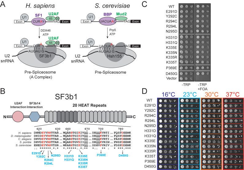
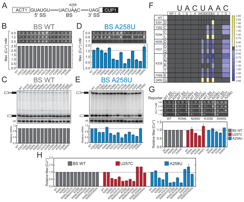
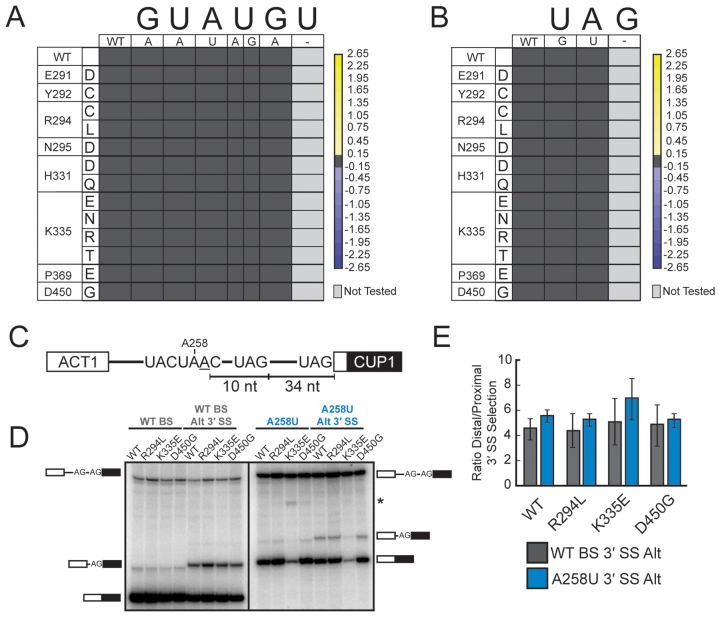
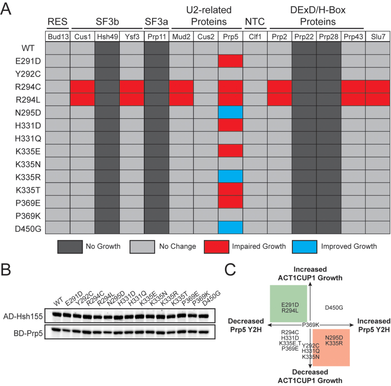
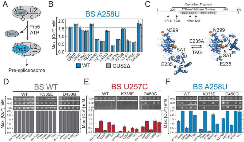
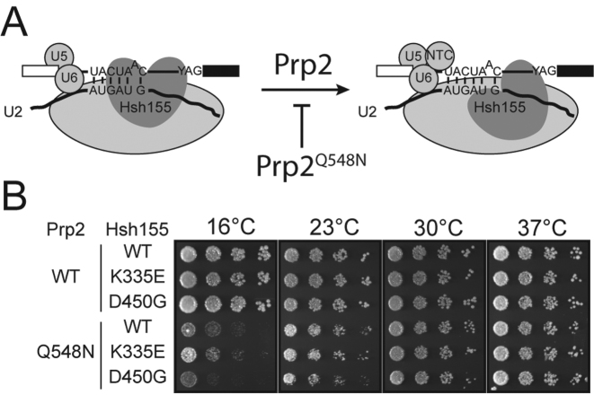
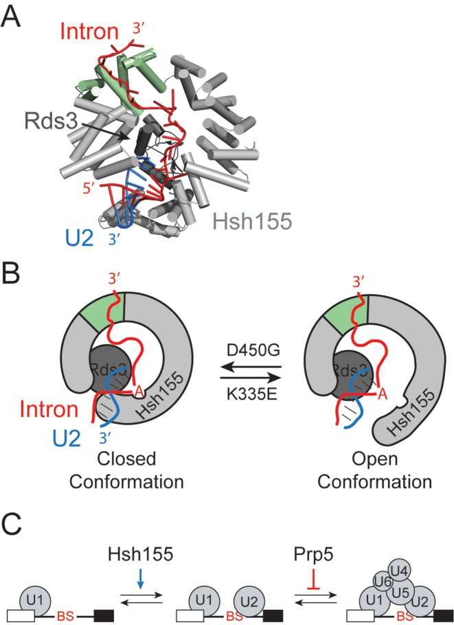
RNA and protein components of the spliceosome work together to identify the 5΄ splice site, the 3΄ splice site, and the branchsite (BS) of nascent pre-mRNA. SF3b1 plays a key role in recruiting the U2 snRNP to the BS. Mutations in human SF3b1 have been linked to many diseases such as myelodysplasia (MDS) and cancer. We have used SF3b1 mutations associated with MDS to interrogate the role of the yeast ortholog, Hsh155, in BS selection and splicing. These alleles change how the spliceosome recognizes the BS and alter splicing when nonconsensus nucleotides are present at the -2, -1 and +1 positions relative to the branchpoint adenosine. This indicates that changes in BS usage observed in humans with SF3b1 mutations may result from perturbation of a conserved mechanism of BS recognition. Notably, different HSH155 alleles elicit disparate effects on splicing: some increase the fidelity of BS selection while others decrease fidelity. Our data support a model wherein conformational changes in SF3b1 promote U2 association with the BS independently of the action of the DEAD-box ATPase Prp5. We propose that SF3b1 functions to stabilize weak U2/BS duplexes to drive spliceosome assembly and splicing.


Figures

Figure 1.
Figure 1.
MDS alleles of Hsh155 do not affect proliferation in yeast. (A) Schematic comparison of pre-spliceosome formation in S. cerevisiae and H. sapiens. Hsh155/SF3b1 function as part of the U2 snRNP, interacting with the BS/U2 snRNA duplex and downstream intronic RNA. (B) (Top) Schematic primary structure of SF3b1, with regions known to interact with other splicing factors indicated. (Bottom) Alignment of sequences from H. sapiens, D. melanogaster, C. elegans, S. pombe and S. cerevisae. Positions found to be frequently mutated in MDS and CLL are shown in red and the amino acid numbering corresponds to H. sapiens SF3b1. The most frequently occurring mutations at those positions are shown in blue with the numbering for S. cerevisiae Hsh155. (C) Haploid yeast expressing only HSH155MDS alleles are viable when plated on FOA. (D) Representative temperature sensitivity growth assays of Hsh155MDS strains plated on YPD. No growth defects are observed in haploid strains expressing only Hsh155MDS plated on YPD at 16, 25, 30 or 37°C. Successive 10-fold dilutions of a OD600 = 0.5 culture are shown.
Figure 2.
Figure 2.
MDS mutations alter the splicing of introns with nonconsensus BS sequences. (A) Schematic representation of the ACT1-CUP1 reporter pre-mRNA. The consensus sequences of the yeast 5΄ SS, BS, and 3΄ SS are shown. The position of A258 is noted and the branchpoint adenosine is underlined. (B) Cu2+ growth assay of strains carrying an ACT1-CUP1 reporter plasmid with a consensus intron. Representative images are shown at the top and the maximum [Cu2+] at which growth was observed is plotted below. (C) Determination of ACT1-CUP1 reporter RNA levels by primer extension from isolated total yeast RNA. (Top) Positions of the pre-mRNA and mRNA are noted in the primer extension polyacrylamide gel. (Middle) Primer extension analysis of the U6 snRNA was used as an internal control and analyzed on the same gel as shown in the top panel. (Bottom) Quantification of the amount of ACT1-CUP1 mRNA after normalization to U6 for each strain. U6 bands are taken from the same gel and contrast has been adjusted. (D) Cu2+ growth assay of strains carrying an ACT1-CUP1 reporter plasmid with a A258U nonconsensus BS. (E) Determination of A258U ACT1-CUP1 reporter RNA levels by primer extension from isolated total yeast RNA. (F) Heatmap summarizing mutant ACT1-CUP1 reporter data for all BS reporters tested. Plotted data represent the log2 transform of the ratio of the maximum [Cu2+] at which growth was observed for the indicated Hsh155MDS mutant to the maximum [Cu2+] at which growth was observed for Hsh155WT. Purple colors indicate decreased growth relative to Hsh155WT, and yellow colors indicate improved growth. (G) Cu2+ growth assay of merodiploid strains expressing the indicated HSH155MDS allele from a plasmid in addition to the chromosomal copy of Hsh155WT for the WT, U257C and A258U ACT1-CUP1 splicing reporters. (H) Cu2+ growth assay of strains expressing Hsh155 proteins harboring multiple MDS mutations for the WT, U257C, and A258U ACT1-CUP1 splicing reporters. In panels B, D-E, and G-H, each bar represents the average of three independent experiments, and error bars represent the standard deviation.
Figure 3.
Figure 3.
MDS mutations do not affect the splicing of introns containing nonconsensus 5΄ SS and 3΄ SS or 3΄ SS selection. (A) Heatmap summarizing mutant ACT1-CUP1 reporter data for all 5΄ SS substitution reporters tested. Data were normalized and the heatmap generated as in Figure 2F. No changes in 5΄ SS usage were observed. (B) Heatmap summarizing mutant ACT1-CUP1 reporter data for all 3΄ SS substitution reporters tested. Data were normalized and the heatmap generated as in Figure 2F. No changes in 3΄ SS usage were observed. (C) Schematic representation of the ACT1-CUP1 reporters used to evaluate cryptic 3΄ SS selection. The cryptic 3΄ SS is located 10 nt downstream of the branchpoint adenosine and 34 nt upstream of the canonical 3΄ SS. Reporters containing both a consensus BS and an A258U substitution were used. (D) Primer extension and PAGE analysis of spliced products of the ACT1-CUP1 reporters shown in (C) from total RNA isolated from the given yeast strains. Positions of the pre-mRNA and mRNA products are noted. The reporter containing the A258U nonconsensus BS also contains a larger 3΄ exon leading to shift in electrophoretic mobility between the consensus and nonconsensus reporter RNAs. The asterisk (*) indicates an unknown band that was not reproducible. (E) Quantification of the data shown in (D) for 3΄ SS usage by the Hsh155WT and given Hsh155MDS strains. Bars represent the average of three independent experiments, and error bars represent the standard deviation.
Figure 4.
Figure 4.
MDS mutations perturb interactions between Hsh155 and Prp5 but leave most other interactions intact. (A) Pseudo-heatmap showing the observed Y2H interactions of Hsh155 upon incorporation of MDS mutations with the given splicing factors. Red indicates an impaired growth relative to Hsh155WT when plated on media that is selective for the Y2H interaction (-His dropout media), blue indicates improved growth, and light grey indicates no change. Dark grey indicates no observable Y2H interaction. (B) Representative western blot confirming expression of the fusion proteins to HSH155MDS used in the Y2H assay. Expression of each potential interacting partner was also confirmed by western blotting and expression of Prp5 is also shown as a representative example. (C) Graphical representation of the relationship between changes in yeast growth observed with the BS A258U ACT1-CUP1 splicing reporter (Figure 2D) and altered interactions observed by Y2H (Figure 4A). Shaded areas represent predictions made from a previously described model for Prp5-based BS fidelity in which retention of Prp5 leads to increased fidelity (red) and weakening of the Prp5 interaction leads to relaxed fidelity (green) (37).
Figure 5.
Figure 5.
Hsh155 MDS mutants affect BS fidelity at a different step than Prp5 proofreading. (A) Cartoon depicting the proposed ATP-dependent function of Prp5 in displacement of Cus2 from the U2 snRNP during spliceosome assembly. (B) Comparison of Cu2+ growth assays using the nonconsensus A258U BS substitution ACT1-CUP1 reporter pre-mRNA between strains containing and lacking the Cus2 protein. No significant differences were observed between the two strains. (C) (Top) Schematic of Prp5 structure indicating the positions of Prp5 mutations used in this assay relative to the two DEAD-box RecA-like domains and the fragment of Prp5 whose structure has been determined by X-ray crystallography. (Bottom) Prp5 has been proposed to undergo a conformational change to promote splicing. The open structure (left) represents the structure determined by X-ray crystallography (pdb 4LJY) while the closed structure (right) is believed to be necessary for ATP hydrolysis and was modeled based on structures of other DEAD-box proteins (coordinates for the closed structure were obtained from Yong-Zhen Xu and Charles Query) (53). Positions of the Prp5 mutations used in this study are noted. The E235A mutation is believed to favor the closed conformation while the TAG mutation of the SAT-motif is believed to favor the open conformation. It is unclear if the N399D mutation used here would impact conformational switching. (D) Cu2+ growth assay for strains containing the Hsh155 WT, K335E, or D450G alleles in combination with the given Prp5 mutations (see text for additional explanation of each Prp5 mutation) with the ACT1-CUP1 reporter containing a consensus BS. (E) Cu2+ growth assay for combinations of Hsh155 and Prp5 as in part (D) except the U257C nonconsensus BS ACT1-CUP1 reporter was used. (F) Cu2+ growth assay for combinations of Hsh155 and Prp5 as in part (D) except the A258U nonconsensus BS ACT1-CUP1 reporter was used. In panels B and D–F, bars represent the average of three independent experiments, and error bars represent the standard deviation.
Figure 6.
Figure 6.
MDS mutations interact genetically with a Prp2 mutation. (A) Cartoon schematic of Prp2-dependent activation of the spliceosome. Prp2 is believed to destabilize Hsh155 as well as the rest of the SF3b complex from interacting with the BS. The PRP2Q548N allele likely stalls this process at low temperatures (34). (B) Representative temperature sensitivity growth assays of the given Hsh155 variants in combination with Prp2WT or Prp2Q548N when plated on YPD at the given temperatures. Hsh155K335E partially suppresses Prp2Q548N and Hsh155D450G enhances cold sensitivity.
Figure 7.
Figure 7.
Models for SF3b1/Hsh155 function in BS duplex stabilization during splicing. (A) Cartoon representation of Hsh155 (light grey and green) and Rds3 (dark gray) bound to the U2 snRNA/BS duplex from the cryo-EM structure of the yeast Bact spliceosome (25). The region of Hsh155 containing the MDS mutations studied here is shown in green. (B) Model for SF3b1/Hsh155 action at the BS. In addition to the structure shown in (A), Hsh155 must also exist in a conformation that permits release of the U2 snRNA/BS RNA duplex and splicing. MDS mutations impact Hsh155 conformation and lead to changes that affect recognition and stabilization of the BS duplex. Mutations that increase splicing of nonconsensus BS (e.g. Hsh155D450G) could stabilize the ‘closed’ or BS duplex bound form whereas mutations that inhibit splicing (e.g. Hsh155K335E) could favor an open form that is necessary for splicing catalysis but does not help stabilize a mismatched duplex during spliceosome assembly. (C) Model for opposing activities of SF3b1/Hsh155 and Prp5 during splicing. SF3b1 functions to stabilize U2 snRNA/BS duplex formation, particularly at nonconsensus or weak BS. Prp5 proofreading opposes this function to enforce BS fidelity by blocking tri-snRNP association. The relative activities of SF3b1/Hsh155 and Prp5 at particular BS may be used to promote or inhibit spliceosome assembly.

Similar articles

Cited by

References

    1. Anczuków O., Krainer A.R.. Splicing-factor alterations in cancers. RNA. 2016; 22:1285–1301. - PMC - PubMed
    1. Dvinge H., Kim E., Abdel-Wahab O., Bradley R.K.. RNA splicing factors as oncoproteins and tumour suppressors. Nat. Rev. Cancer. 2016; 16:413–430. - PMC - PubMed
    1. Coovert D.D., Le T.T., McAndrew P.E., Strasswimmer J., Crawford T.O., Mendell J.R., Coulson S.E., Androphy E.J., Prior T.W., Burghes A.H.. The survival motor neuron protein in spinal muscular atrophy. Hum. Mol. Genet. 1997; 6:1205–1214. - PubMed
    1. McKie A.B., McHale J.C., Keen T.J., Tarttelin E.E., Goliath R., van Lith-Verhoeven J.J., Greenberg J., Ramesar R.S., Hoyng C.B., Cremers F.P. et al. . Mutations in the pre-mRNA splicing factor gene PRPC8 in autosomal dominant retinitis pigmentosa (RP13). Hum. Mol. Genet. 2001; 10:1555–1562. - PubMed
    1. Vithana E.N., Abu-Safieh L., Allen M.J., Carey A., Papaioannou M., Chakarova C., Al-Maghtheh M., Ebenezer N.D., Willis C., Moore A.T. et al. . A human homolog of yeast pre-mRNA splicing gene, PRP31, underlies autosomal dominant retinitis pigmentosa on chromosome 19q13.4 (RP11). Mol. Cell. 2001; 8:375–381. - PubMed

MeSH terms