. 2017 Dec;3(1):9.
doi: 10.1186/s40792-016-0284-9. Epub 2017 Jan 6.

Undifferentiated carcinoma of the ampulla of Vater

Affiliations

Undifferentiated carcinoma of the ampulla of Vater

Misuzu Yamada et al. Surg Case Rep. 2017 Dec.

Abstract

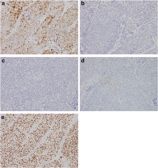
Undifferentiated carcinoma of the ampulla of Vater is a rare disease with unclear and clinical characteristics and prognosis. Here, we report the case of a 61-year-old man with undifferentiated carcinoma of the ampulla of Vater. He presented to our hospital with an increase in hepatobiliary system enzymes that was detected during a health check-up. Imaging and endoscopy demonstrated a tumor with ulcer in the ampulla of Vater, which was diagnosed as a carcinoma by biopsy. No distant metastasis was observed. Subtotal stomach-preserving pancreaticoduodenectomy was performed. Undifferentiated carcinoma was confirmed based on the presence of small round atypical cells with the formation of a solid alveolar lesion on histopathological examination and immunohistochemical staining that was positive for CAM 5.2 but negative for chromogranin A and synaptophysin. The tumor infiltrated the duodenum, but not the pancreas; no lymph node metastasis was observed. However, liver metastases were detected 2 months postoperatively. Chemotherapy was performed, and the tumor size temporality decreased; however, it grew in size again, and the patients subsequently died of the primary disease 15 months postoperatively. Undifferentiated carcinoma of the ampulla of Vater is a very rare histological type. More number of cases is necessary to clarify optimal treatment.

Keywords: Ampulla of Vater; Cisplatin; Gemcitabine; Pancreaticoduodenectomy; Undifferentiated carcinoma.


Figures

Fig. 1
Fig. 1
Abdominal CT. Axial (a) and coronal (b) images show a 3-cm tumor in the ampulla on Vater (white arrow) and dilatation of the common bile duct
Fig. 2
Fig. 2
Upper gastrointestinal endoscopy. An ulcerative tumor with raised margins in the ampulla of Vater
Fig. 3
Fig. 3
Gross findings of the resected specimen. The resected specimens demonstrated an ulcerative tumor with raised margins (a) and involvement of the inferior bile duct (b)
Fig. 4
Fig. 4
Pathological examination of the resected specimen. The tumor is highly cellular with minimal stroma and small cells with scant cytoplasm arranged in solid nests (Hematoxylin and Eosin stain, ×200)
Fig. 5
Fig. 5
Transversely cut surface of the resected specimen. The small round atypical cells without ductal differentiation are present in the majority of the tumor (parts surrounded by solid line). Ductal component is found in the small portion facing the lumen of the duodenum (part surrounded by dotted line)
Fig. 6
Fig. 6
Immunohistochemical staining of the resected specimen. The tumor is positive for CAM 5.2 (a) but negative for synaptophysin (b) and chromogranin A (c). CD56 is weakly positive (d). The labeling index of MIB-1 was >90% (e) (All photomicrographs, ×200)
Fig. 7
Fig. 7
Serial abdominal computed tomography findings. Two months postoperatively, liver metastases are demonstrated (a), which increased 6 months postoperatively (b). The liver metastases resolved after 2 months of restarting gemcitabine plus cisplatin (c), but it enlarged again 2 months later (d)

References

    1. Guo KJ, Yamaguchi K, Enjoji M. Undifferentiated carcinoma of the gallbladder. A clinicopathologic, histochemical, and immunohistochemical study of 21 patients with a poor prognosis. Cancer. 1988;61:1872–9. doi: 10.1002/1097-0142(19880501)61:9<1872::AID-CNCR2820610925>3.0.CO;2-Q. - DOI - PubMed
    1. Park HJ, Jang KT, Choi DW, Heo JS, Choi SH. Clinicopathologic analysis of undifferentiated carcinoma of the gallbladder. J Hepatobiliary Pancreat Sci. 2014;21:58–63. doi: 10.1002/jhbp.3. - DOI - PubMed
    1. Fujikawa T, Tanaka A, Abe T, Yoshimoto H, Tokumitsu Y, Tada S, et al. Undifferentiated carcinoma of the common bile duct with intraductal tumor thrombi: report of a case. Surg Today. 2011;41:579–84. doi: 10.1007/s00595-009-4304-2. - DOI - PubMed
    1. Sato T, Yamamoto K, Ouchi A, Imaoka Y, Tokumura H, Matsushiro T. Undifferentiated carcinoma of the duodenal ampulla. J Gastroenterol. 1995;30:517–9. doi: 10.1007/BF02347570. - DOI - PubMed
    1. Albores-Saavedra J, Hruban RH, Klimstra DS, Zamboni G. Invasive adenocarcinoma of the ampullary region. In: Bosman FT, Carneiro F, Hruban RH, Theise ND, editors. WHO Classification of Tumours of the Digestive System. 4. Lyon: International Agency for Research on Cancer; 2010. pp. 87–91.

LinkOut - more resources