. 2017 Feb 10:247:153-166.
doi: 10.1016/j.jconrel.2016.12.038. Epub 2017 Jan 4.

An intraocular drug delivery system using targeted nanocarriers attenuates retinal ganglion cell degeneration

Affiliations

An intraocular drug delivery system using targeted nanocarriers attenuates retinal ganglion cell degeneration

Lei Zhao et al. J Control Release. .

Abstract

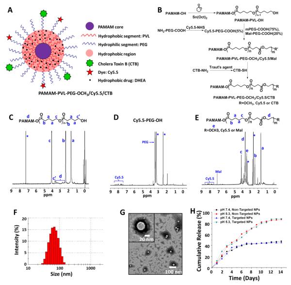
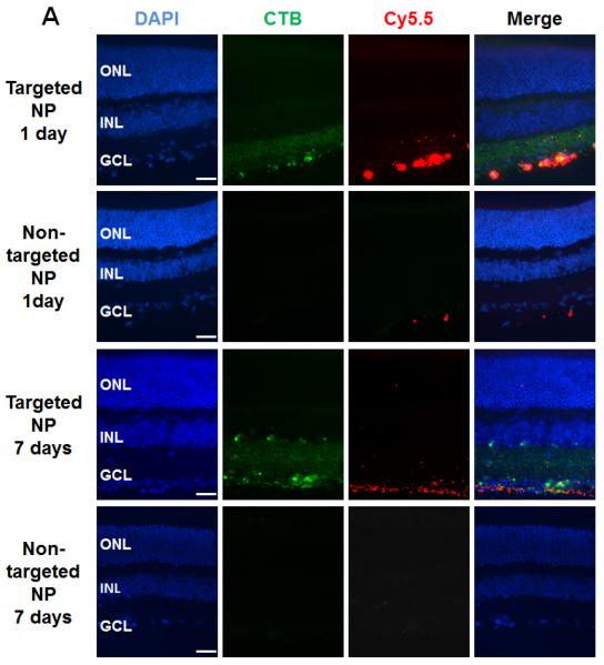
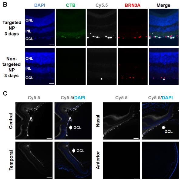
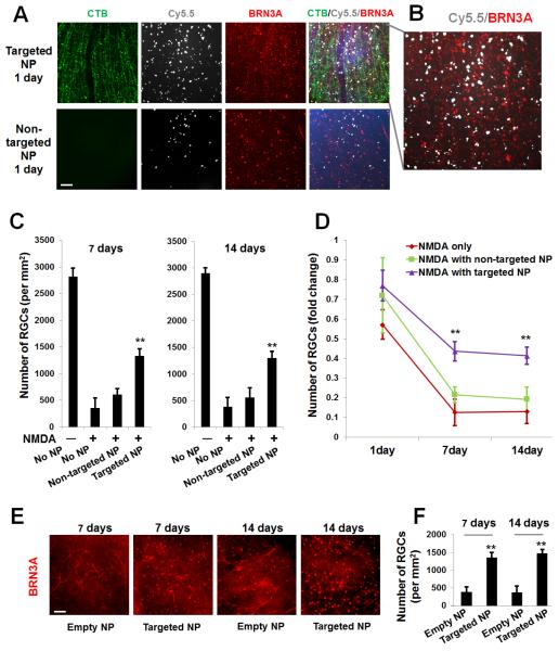
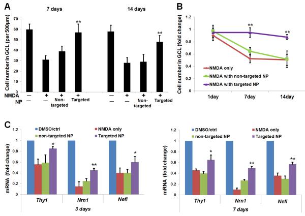
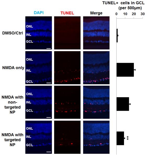
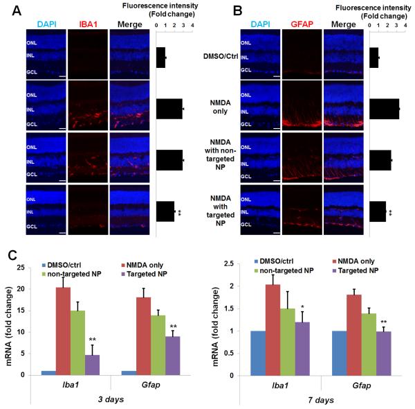
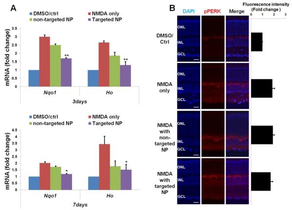
Glaucoma is a common blinding disease characterized by loss of retinal ganglion cells (RGCs). To date, there is no clinically available treatment directly targeting RGCs. We aim to develop an RGC-targeted intraocular drug delivery system using unimolecular micelle nanoparticles (unimNPs) to prevent RGC loss. The unimNPs were formed by single/individual multi-arm star amphiphilic block copolymer poly(amidoamine)-polyvalerolactone-poly(ethylene glycol) (PAMAM-PVL-PEG). While the hydrophobic PAMAM-PVL core can encapsulate hydrophobic drugs, the hydrophilic PEG shell provides excellent water dispersity. We conjugated unimNPs with the cholera toxin B domain (CTB) for RGC-targeting and with Cy5.5 for unimNP-tracing. To exploit RGC-protective sigma-1 receptor (S1R), we loaded unimNPs with an endogenous S1R agonist dehydroepiandrosterone (DHEA) as an FDA-approved model drug. These unimNPs produced a steady DHEA release in vitro for over two months at pH7.4. We then co-injected (mice, intraocular) unimNPs with the glutamate analog N-methyl-d-aspartate (NMDA), which is excito-toxic and induces RGC death. The CTB-conjugated unimNPs (i.e., targeted NPs) accumulated at the RGC layer and effectively preserved RGCs at least for 14days, whereas the unimNPs without CTB (i.e., non-targeted NPs) showed neither accumulation at nor protection of NMDA-treated RGCs. Consistent with S1R functions, targeted NPs relative to non-targeted NPs showed markedly better inhibitory effects on apoptosis and oxidative/inflammatory stresses in the RGC layer. Hence, the DHEA-loaded, CTB-conjugated unimNPs represent an RGC/S1R dual-targeted nanoplatform that generates an efficacious template for further development of a sustainable intraocular drug delivery system to protect RGCs, which may be applicable to treatments directed at glaucomatous pathology.

Keywords: Cholera toxin B domain; Excitotoxicity; Ganglion cell targeting; Retina; Sigma-1 receptor; Targeted drug delivery; Unimolecular micelles.


Figures

Figure 1
Figure 1. Synthesis and characterization of targeted and non-targeted unimNPs
(A) A schematic illustration of multifunctional unimolecular micelle nanoparticles formed by multi-arm star amphiphilic block copolymer PAMAM–PVL–PEG–Cy5.5/CTB for targeted delivery of DHEA to RGCs. (B) Synthesis scheme of multi-arm star amphiphilic block copolymer PAMAM–PVL–PEG–Cy5.5/CTB. 1H NMR spectra of (C) PAMAM–PVL–OH, (D) Cy5.5–PEG–COOH, and (E) PAMAM–PVL–PEG–OCH3/Cy5.5/Mal. (F) DLS analysis and (G) TEM images of the unimNPs. (H) In vitro DHEA release profiles from DHEA-loaded, non-targeted and targeted unimNPs at two different pHs.
Figure 2
Figure 2. Accumulation of targeted NPs at the mouse RGC layer after intravitreal injection
Targeted NPs (i.e., DHEA-loaded CTB-unimNPs) and non-targeted NPs control (i.e., DHEA-loaded unimNPs), both mixed with NMDA, were injected respectively into the right eye (or left eye, randomly assigned) of two separate groups of mice,. Each injection (total 2 μl) contained 0.5 μg DHEA, 40 mM NMDA, and 2 μg unimNP. At 1, 3, and 7 days after injection, mice were euthanized and retinal sections prepared for fluorescence microscopy. Retinal layers were distinguished by DAPI staining: ONL, outer nuclear layer; INL, inner nuclear layer; GCL, retinal ganglion cell (RGC) layer. (A) Localization of NP (Cy5.5-conjugated, red). While targeted NPs were accumulated at the RGC layer, non-targeted NPs were barely detectable. CTB was labeled green (FITC). (B) Co-localization of NP (white, pseudo-color) with RGCs, which were detected by immunostaining of the marker protein BRN3A (red). (C) Overview of the entire mouse eye section showing accumulation of targeted NPs (white, pseudo-color) along GCL in the retina. The anterior image shows the cornea which is Cy5.5 negative. Scale bar: A and B, 50 μm; C, 200 μm.
Figure 3
Figure 3. Rescue of RGCs by targeted NPs following intravitreal injection (counting on whole-mounts)
Intravitreal injections were performed as described in Figure 2. Mice were euthanized at indicated time points and retinal whole mounts were prepared for fluorescence microscopy. (A) Representative images showing distribution of CTB (green), Cy5.5 (white), and BRN3A-positive nuclei (red). Scale bar: 100 μm. (B). Enlarged image in A showing Cy5.5 and BRN3A. (C). Quantification of BRN3A-positive cells: mean ± SE; n = 7–10 animals. **P < 0.01, compared to NMDA only (no NPs). (D). Data are re-plotted as time course of nuclei number fold change versus vehicle (DMSO) control. **P < 0.01 compared to NMDA only (no NPs). The data show that while targeted NPs preserved RGC layer cells, non-targeted NPs had no significant effect. (E) Representative images showing BRN3A-positive nuclei of retinal whole mounts collected at 7 and 14 days after injection of CTB-unimNPs without (empty NP) or with (targeted NP) DHEA loaded (the same amount as in A). Scale bar: 100 μm. (F). Quantification of BRN3A-positive cells in E: mean ± SE; n = 7–10 animals. **P < 0.01, compared to empty NPs.
Figure 4
Figure 4. Rescue of RGCs by targeted NPs following intravitreal injection (counting on vertical sections)
Injections were performed as described in Figure 2. Eyeballs were collected at indicated time points after injection for the preparation of retinal sections (nuclei counting) or homogenates (RT-PCR). Quantification: mean ± SE; n = 7–10 animals. (A) The number of nuclei (per 500 μm retina length) was counted at the RGC layer. **P < 0.01, compared to NMDA only (no NPs). (B) Data are re-plotted as time course of nuclei number fold change versus vehicle (DMSO) control. **P < 0.01 compared to NMDA only (no NPs). The data show that while targeted NPs preserved RGC layer cells, non-targeted NPs had no significant effect. (C) Expression of RGC marker genes was determined by qRT-PCR. **P < 0.01, *P < 0.05, compared to NMDA only.
Figure 5
Figure 5. Prevention of NMDA-induced increase of TUNEL-positive cells by targeted NPs following intravitreal injection
Injections were performed as described in Figure 2. Eyeballs were collected at 1 day after injection. TUNEL staining was performed on retinal cryosections. Scale Bar: 50 μm. Quantification: mean ± SE of TUNEL-positive nuclei at the RGC layer (per 500 μm retina length); n > 5 animals; **P < 0.01 compared to NMDA alone.
Figure 6
Figure 6. Inhibition of NMDA-induced activation of retinal microglia and macroglia by targeted NPs following intravitreal injection
Injections were performed as described in Figure 2. Eyeballs were collected for preparation of retinal cryosections (immunohistochemistry) or homogenates (qRT-PCR). Quantification: mean ± SE; normalization to vehicle control; n > 5 animals; **P < 0.01, *P < 0.05, compared to NMDA only. Immunostaining of (A) IBA1 or (B) GFAP were performed on retinal sections collected 3 days after injection and the area including INL and GCL was used for quantification. Scale bar: 50 μm. (C) Levels of mRNA of Iba1 and Gfap were determined by qRT-PCR using unfixed samples collected at 3 or 7 days after injection.
Figure 7
Figure 7. Reduction of NMDA-induced oxidative stress by targeted NPs following intravitreal injection
Injections were performed as described in Figure 2. Eyeballs were collected for preparation of retinal cryosections (immunohistochemistry) or homogenates (RT-PCR). Quantification: mean ± SE; normalization to vehicle control; n > 5 animals; **P < 0.01, *P < 0.05, compared to NMDA only. (A) Levels of mRNA of Nqo1 and Ho1 were determined by RT-PCR using unfixed samples collected at 3 or 7 days after injection. (B) Immuno-staining of pPERK performed on retinal sections collected at 3 days after injection and the area including INL and GCL was used for quantification. Scale bar: 50 μm.

References

    1. Nickells RW, Howell GR, Soto I, John SW. Under pressure: cellular and molecular responses during glaucoma, a common neurodegeneration with axonopathy. Annual review of neuroscience. 2012;35:153–179. - PubMed
    1. Martin PM, Ola MS, Agarwal N, Ganapathy V, Smith SB. The sigma receptor ligand (+)-pentazocine prevents apoptotic retinal ganglion cell death induced in vitro by homocysteine and glutamate, Brain research. Molecular brain research. 2004;123:66–75. - PMC - PubMed
    1. Dun Y, Thangaraju M, Prasad P, Ganapathy V, Smith SB. Prevention of excitotoxicity in primary retinal ganglion cells by (+)-pentazocine, a sigma receptor-1 specific ligand. Investigative ophthalmology & visual science. 2007;48:4785–4794. - PMC - PubMed
    1. Ha Y, Dun Y, Thangaraju M, Duplantier J, Dong Z, Liu K, Ganapathy V, Smith SB. Sigma receptor 1 modulates endoplasmic reticulum stress in retinal neurons. Investigative ophthalmology & visual science. 2011;52:527–540. - PMC - PubMed
    1. Tchedre KT, Huang RQ, Dibas A, Krishnamoorthy RR, Dillon GH, Yorio T. Sigma-1 receptor regulation of voltage-gated calcium channels involves a direct interaction. Investigative ophthalmology & visual science. 2008;49:4993–5002. - PubMed

MeSH terms