. 2017 Jul 1;32(7):1137-1145.
doi: 10.1093/ndt/gfw393.

Acute genetic ablation of pendrin lowers blood pressure in mice

Affiliations

Acute genetic ablation of pendrin lowers blood pressure in mice

Francesco Trepiccione et al. Nephrol Dial Transplant. .

Abstract

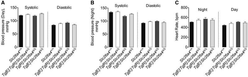
Background: Pendrin, the chloride/bicarbonate exchanger of β-intercalated cells of the renal connecting tubule and the collecting duct, plays a key role in NaCl reabsorption by the distal nephron. Therefore, pendrin may be important for the control of extracellular fluid volume and blood pressure.

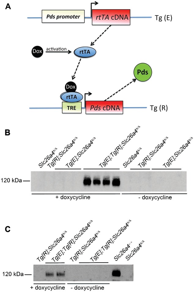
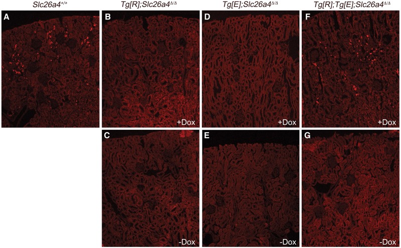
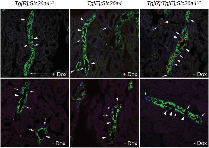
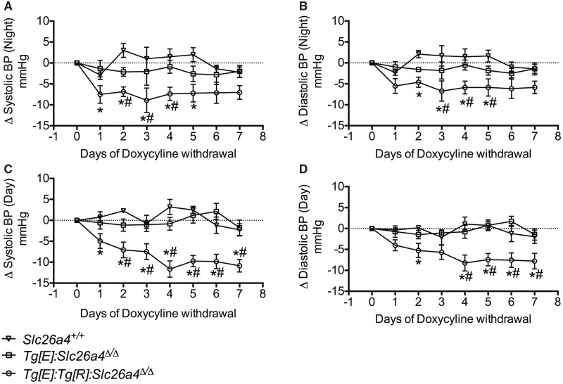
Methods: Here, we have used a genetic mouse model in which the expression of pendrin can be switched-on in vivo by the administration of doxycycline. Pendrin can also be rapidly removed when doxycycline administration is discontinued. Therefore, our genetic strategy allows us to test selectively the acute effects of loss of pendrin function.

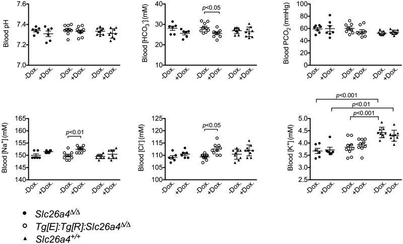
Results: We show that acute loss of pendrin leads to a significant decrease of blood pressure. In addition, acute ablation of pendrin did not alter significantly the acid-base status or blood K + concentration.

Conclusion: By using a transgenic mouse model, avoiding off-target effects related to pharmacological compounds, this study suggests that pendrin could be a novel target to treat hypertension.

Keywords: chloride; diuretics; hypertension; intercalated cells; pendrin.


Figures

FIGURE 1
FIGURE 1
Schematic representation of the transgenes and its validation. (A) The transgenes are crossed onto a Slc26a4Δ/Δ background so the transgene Tg[R] is the only source of pendrin. When mice are administrated with doxycycline, rtTA expression is driven by the pendrin promoter and cis-regulatory elements in mice that normally express pendrin (i.e. the non-β-intercalated cells in the kidney), and then pendrin mRNA expression stops when doxycycline is withdrawn. Pds, pendrin; rtTA, reverse tetracyclin-dependent transactivator; Dox, doxycycline hyclate; TRE, tetracyclin responsive element. (B and C) Analyses of mouse pendrin expression by immunoblots of membrane fractions from Slc26a4+/+, Slc26a4Δ/Δ mice Tg[R];Slc26a4Δ/Δ, Tg[E];Slc26a4Δ/Δ and Tg[E];Tg[R];Slc26a4Δ/Δ. For the samples labeled ‘ + doxycycline’ tissue was extracted from the renal cortex doxycycline-administered mice for 2–3 weeks in the drinking water. For ‘−doxycycline’ tissue was extracted 5 days after doxycycline administration. Immunoblots were probed with the anti-mouse pendrin antibody.
FIGURE 2
FIGURE 2
Immunolocalization of pendrin on kidney sections from Slc26a4+/+, Tg[R];Slc26a4Δ/Δ, Tg[E];Slc26a4Δ/Δ and Tg[E];Tg[R];Slc26a4Δ/Δ mice. Anti-pendrin antibody stained a subset of renal epithelial cells in the renal cortex of Slc26a4+/+ mice. The same pattern of expression of pendrin was observed in Tg[E];Tg[R];Slc26a4Δ/Δ mice treated for at least 15 days with doxycycline. After 5 days of doxycycline withdrawal, pendrin expression was completely undetectable. As expected, neither Tg[R];Slc26a4Δ/Δ nor Tg[E];Slc26a4Δ/Δ mice expressed pendrin protein, either in the presence or absence of doxycycline treatment.
FIGURE 3
FIGURE 3
Cellular composition of collecting duct from Tg[E];Tg[R];Slc26a4Δ/Δ, Tg[E];Slc26a4Δ/Δ or Tg[R]; Slc26a4Δ/Δ mice. Colocalization studies with anti-pendrin (red), anti-AE1 (blue) and anti-AQP2 (green) antibodies. Doxycycline administration for 2 weeks does not induce pendrin staining in Tg[R];Slc26a4Δ/Δ or Tg[E];Slc26a4Δ/Δ mice, while the expression of pendrin at the apical pole of a subset of cells in cortical renal tubules that are also labeled by Ae1- and Aqp2-specific antibodies, and thus are identified as connecting tubules or cortical collecting ducts, occurs in Tg[E];Tg[R];Slc26a4Δ/Δ mice. Pendrin staining is restricted to a subset of cells that are identified as β-intercalated cells because they are negative for Ae1 or for Aqp2 staining, and it disappears in mice in which doxycycline has been withdrawn for 5 days.
FIGURE 4
FIGURE 4
Blood gas analysis and electrolytes concentration in Slc26a4+/+, Slc26a4Δ/Δ and Tg[E];Tg[R];Slc26a4Δ/Δ mice during doxycycline administration or after doxycycline withdrawal. Mice that do not expressed pendrin have significantly higher blood [HCO3] and lower blood [Na+] and [Cl]. However, the changes were of limited amplitude (<4 mmol/L). Statistical significance was tested by ANOVA followed by Sidak’s post hoc test for multiple comparisons (n=6, 9 and 9, for Slc26a4+/+, Slc26a4Δ/Δ and Tg[E];Tg[R];Slc26a4Δ/Δ mice, respectively).
FIGURE 5
FIGURE 5
(A and B) Blood pressure and (C) heart rate in basal conditions in Slc26a4+/+, Tg[E];Tg[R];Slc26a4Δ/Δ Tg[R];Slc26a4Δ/Δ, Tg[E];Slc26a4Δ/Δ and Slc26a4Δ/Δ mice. Systolic and diastolic blood pressures were continuously recorded by radiotelemetry. During all the experiments, mice were fed a normal salt (0.3% Na+) diet and were administrated doxycycline during at least 2–3 weeks. Data are presented as the mean ± standard error of the values obtained during either the active (night) or inactive (day) period; n = 4–6 per group. The differences in heart rate, systolic and diastolic blood pressures were tested by one-way ANOVA.
FIGURE 6
FIGURE 6
Effects of doxycycline withdrawal on the time course of (A and C) systolic (B and D) and diastolic blood pressures of Slc26a4+/+, Tg[E];Tg[R];Slc26a4Δ/Δ, Tg[E];Slc26a4Δ/Δ mice. Systolic and diastolic blood pressures were continuously recorded by radiotelemetry. During all the experiments, mice were fed a normal salt (0.3% Na+) diet. Mice were administrated doxycycline for at least 2–3 weeks and then the time course of blood pressure was monitored after doxycycline withdrawal. Data are presented as the mean ± standard error of the values obtained during either the active (night) or inactive (day) period; n = 4–6 per group. The differences were tested using a two-way ANOVA for repeated measures followed by Student–Newman–Keuls post hoc test. Both systolic and diastolic blood pressures significantly (P < 0.05) dropped from Day 1 in Tg[E];Tg[R];Slc26a4Δ/Δ mice after doxycycline withdrawal versus the control period (Day 0), while they were not affected in Slc26a4+/+ or Tg[E];Slc26a4Δ/Δ control mice. * indicates statistical significance (P < 0.05 or lower) versus Slc26a4+/+ mice; # indicates statistical significance (P < 0.05 or lower) versus Tg[E];Slc26a4Δ/Δ mice.

Similar articles

Cited by

References

    1. Scott DA, Wang R, Kreman TM. et al. The Pendred syndrome gene encodes a chloride-iodide transport protein. Nat Genet 1999; 21: 440–443 - PubMed
    1. Reardon W, Trembath RC.. Pendred syndrome. J Med Genet 1996; 33: 1037–1040 - PMC - PubMed
    1. Pendred V. Deaf-mutism and goitre. Lancet 1896; 148: 532
    1. Leviel F, Hubner CA, Houillier P. et al. . The Na+-dependent chloride-bicarbonate exchanger slc4a8 mediates an electroneutral Na+ reabsorption process in the renal cortical collecting ducts of mice. J Clin Invest 2010; 120: 1627–1635 - PMC - PubMed
    1. Quentin F, Chambrey R, Trinh-Trang-Tan MM. et al. The Cl-/HCO3- exchanger pendrin in the rat kidney is regulated in response to chronic alterations in chloride balance. Am J Physiol Renal Physiol 2004; 287: F1179–F1188 - PubMed