. 2017 Feb 2;100(2):216-227.
doi: 10.1016/j.ajhg.2016.12.010. Epub 2017 Jan 5.

Mutations in ATP6V1E1 or ATP6V1A Cause Autosomal-Recessive Cutis Laxa

Affiliations

Mutations in ATP6V1E1 or ATP6V1A Cause Autosomal-Recessive Cutis Laxa

Tim Van Damme et al. Am J Hum Genet. .

Erratum in

  • Mutations in ATP6V1E1 or ATP6V1A Cause Autosomal-Recessive Cutis Laxa.
    Van Damme T, Gardeitchik T, Mohamed M, Guerrero-Castillo S, Freisinger P, Guillemyn B, Kariminejad A, Dalloyaux D, van Kraaij S, Lefeber DJ, Syx D, Steyaert W, De Rycke R, Hoischen A, Kamsteeg EJ, Wong SY, van Scherpenzeel M, Jamali P, Brandt U, Nijtmans L, Korenke GC, Chung BHY, Mak CCY, Hausser I, Kornak U, Fischer-Zirnsak B, Strom TM, Meitinger T, Alanay Y, Utine GE, Leung KCP, Ghaderi-Sohi S, Coucke P, Symoens S, De Paepe A, Thiel C, Haack TB, Malfait F, Morava E, Callewaert B, Wevers RA. Van Damme T, et al. Am J Hum Genet. 2020 Aug 6;107(2):374. doi: 10.1016/j.ajhg.2020.07.013. Am J Hum Genet. 2020. PMID: 32763190 Free PMC article. No abstract available.

Abstract

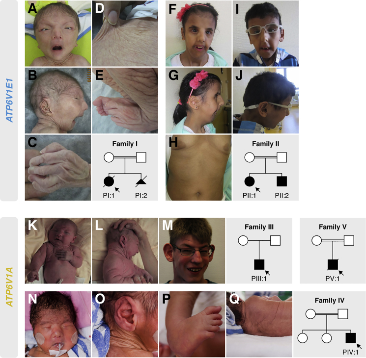
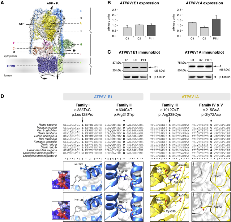
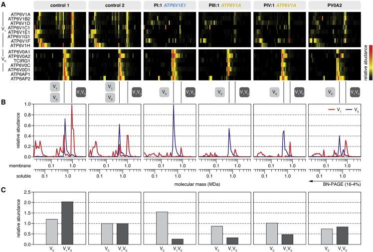
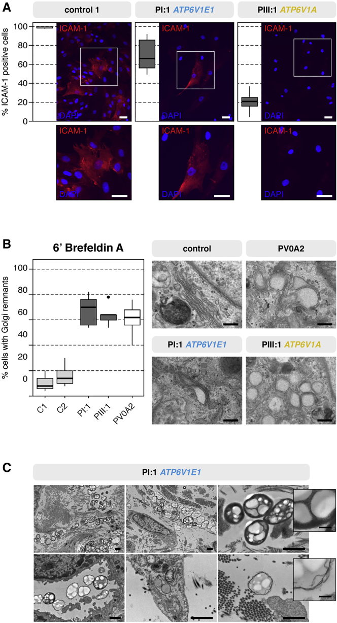
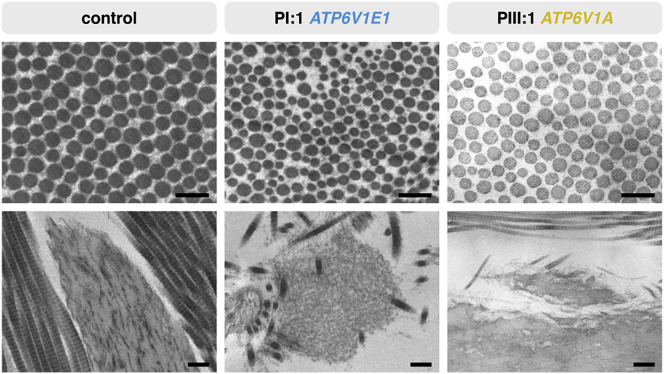
Defects of the V-type proton (H+) ATPase (V-ATPase) impair acidification and intracellular trafficking of membrane-enclosed compartments, including secretory granules, endosomes, and lysosomes. Whole-exome sequencing in five families affected by mild to severe cutis laxa, dysmorphic facial features, and cardiopulmonary involvement identified biallelic missense mutations in ATP6V1E1 and ATP6V1A, which encode the E1 and A subunits, respectively, of the V1 domain of the heteromultimeric V-ATPase complex. Structural modeling indicated that all substitutions affect critical residues and inter- or intrasubunit interactions. Furthermore, complexome profiling, a method combining blue-native gel electrophoresis and liquid chromatography tandem mass spectrometry, showed that they disturb either the assembly or the stability of the V-ATPase complex. Protein glycosylation was variably affected. Abnormal vesicular trafficking was evidenced by delayed retrograde transport after brefeldin A treatment and abnormal swelling and fragmentation of the Golgi apparatus. In addition to showing reduced and fragmented elastic fibers, the histopathological hallmark of cutis laxa, transmission electron microscopy of the dermis also showed pronounced changes in the structure and organization of the collagen fibers. Our findings expand the clinical and molecular spectrum of metabolic cutis laxa syndromes and further link defective extracellular matrix assembly to faulty protein processing and cellular trafficking caused by genetic defects in the V-ATPase complex.

Keywords: ARCL2; ATP6V1A; ATP6V1E1; CDG; Golgi apparatus; V-ATPase; autosomal recessive; cellular trafficking; congenital disorder of glycosylation; cutis laxa.


Figures

Figure 1
Figure 1
Clinical Characteristics and Pedigrees Clinical pictures of PI:1 (at birth, A–E), PII:1 (at age 10 years, F–H), PII:2 (at age 9 years, I and J), PIII:1 (at birth, K and L; at age 15 years, M), and PIV:1 (at birth, N–Q) and pedigrees of all affected families. Clinical pictures of PV:1 were not available.
Figure 2
Figure 2
ATP6V1E1 and ATP6V1A Mutations (A) Alignment of I-Tasser homology models for the human E1 (blue surface shading) and A (yellow surface shading) subunits with a three-dimensional model of the Saccharomyces cerevisiae V-ATPase (PDB: 3J9T). (B) qRT-PCR analysis of ATP6V1E1 and ATP6V1A in cultured fibroblasts from PI:1, PIII:1, and two age- and sex-matched control individuals showed no difference in expression. Data are expressed as the mean, and error bars represent the 95% confidence interval. (C) Immunoblotting against the E1 and A subunits in PI:1, PIII:1, and two age- and sex-matched control individuals showed no difference in protein level in cultured fibroblasts. (D) Clustal Omega protein sequence alignment and structural modeling. The protein sequence of the E1 and A subunits is highly conserved across vertebrates and invertebrates, and all affected amino acid residues are evolutionary highly conserved. Asterisks indicate a single, fully conserved residue, colons indicate strong similar properties (>0.5 in the Gonnet PAM 250 matrix), and periods indicate weak similar properties (≤0.5 in the Gonnet PAM 250 matrix). Structural modeling indicated that all identified mutations affect critical residues within the E1 or A subunit and disrupt inter- or intrasubunit interactions within the V-ATPase complex.
Figure 3
Figure 3
Complexome Profiling (A) Heatmap representations of the migration profiles of the identified V1 and V0 subunits and two V-ATPase assembly factors (ATP6AP1 and ATP6AP2) were created by hierarchical clustering and, for proteins that were not grouped together by the clustering algorithm, by correlation profiling. In control fibroblast cultures, the fully assembled V-ATPase and the separate V1 and V0 domains migrated with apparent molecular masses of 1,000, 600, and 450 kDa, respectively. The majority of V1 subunits were integrated in the complete V-ATPase complex, whereas the vast majority of V0 subunits were integrated in the V0 subassembly. In fibroblast cultures from individuals with ATP6V1E1 or ATP6V1A mutations, the abundance of V1 subunits was markedly lower than in control individuals. (B) Migration profiles of the V1 (red) and V0 (blue) domains show the average value of the abundance of the detected subunits of the respective fraction plotted against the molecular mass. (C) The amount of fully assembled V-ATPase complex was markedly reduced in fibroblast cultures from all individuals with ATP6V1E1 or ATP6V1A mutations, but the amount of V0 domain was unchanged. In PV0A2, an individual with compound-heterozygous ATP6V0A2 mutations, the amount of fully assembled V-ATPase complex was only moderately reduced. To calculate the values, we summarized the abundances of all detected subunits in the respective fraction and normalized them to control 2.
Figure 4
Figure 4
Glycosylation and Vesicular Trafficking Studies (A) Immunocytochemistry of the highly glycosylated ICAM-1 showed a severe reduction in the percentage of ICAM-1-positive fibroblasts. Scale bars represent 25 μm. (B) Retrograde translocation of Golgi membranes to the endoplasmic reticulum was severely delayed in brefeldin-A-treated fibroblasts from PI:1 and PIII:1. Similar to PV0A2 (with ARCL2A), PI:1 and PIII:1 showed a 2- to 3-fold higher percentage of cells retaining Golgi remnants than control individuals. Additionally, TEM showed dilated and fragmented Golgi cisternae in PI:1, PIII:1, and PV0A2. Scale bars represent 250 nm. (C) TEM imaging of the dermis of PI:1 showed the presence of large, heterogeneous vacuolar structures within or near fibroblasts and in the endothelial lumen. Scale bars represent 1 μm or 200 nm (inset).
Figure 5
Figure 5
Ultrastructural Studies of the ECM TEM of the dermis of PI:1 and PIII:1 showed collagen abnormalities with increased interfibrillar space and more variable fibril diameters than did a control sample (top row; scale bars represent 250 nm). Elastic fibers appeared normal in PIII:1 but were very irregular, fragmented, and almost absent in PI:1 (bottom row; scale bars represent 500 nm).

Comment in

References

    1. Forgac M. Vacuolar ATPases: rotary proton pumps in physiology and pathophysiology. Nat. Rev. Mol. Cell Biol. 2007;8:917–929. - PubMed
    1. Cotter K., Stransky L., McGuire C., Forgac M. Recent Insights into the Structure, Regulation, and Function of the V-ATPases. Trends Biochem. Sci. 2015;40:611–622. - PMC - PubMed
    1. Guillard M., Dimopoulou A., Fischer B., Morava E., Lefeber D.J., Kornak U., Wevers R.A. Vacuolar H+-ATPase meets glycosylation in patients with cutis laxa. Biochim. Biophys. Acta. 2009;1792:903–914. - PubMed
    1. Karet F.E., Finberg K.E., Nelson R.D., Nayir A., Mocan H., Sanjad S.A., Rodriguez-Soriano J., Santos F., Cremers C.W.R.J., Di Pietro A. Mutations in the gene encoding B1 subunit of H+-ATPase cause renal tubular acidosis with sensorineural deafness. Nat. Genet. 1999;21:84–90. - PubMed
    1. Kortüm F., Caputo V., Bauer C.K., Stella L., Ciolfi A., Alawi M., Bocchinfuso G., Flex E., Paolacci S., Dentici M.L. Mutations in KCNH1 and ATP6V1B2 cause Zimmermann-Laband syndrome. Nat. Genet. 2015;47:661–667. - PubMed

Publication types

MeSH terms

Substances

Supplementary concepts