. 2017 Feb 16;65(4):743-750.e4.
doi: 10.1016/j.molcel.2016.11.039. Epub 2017 Jan 5.

Initiation of Quality Control during Poly(A) Translation Requires Site-Specific Ribosome Ubiquitination

Affiliations

Initiation of Quality Control during Poly(A) Translation Requires Site-Specific Ribosome Ubiquitination

Szymon Juszkiewicz et al. Mol Cell. .

Abstract

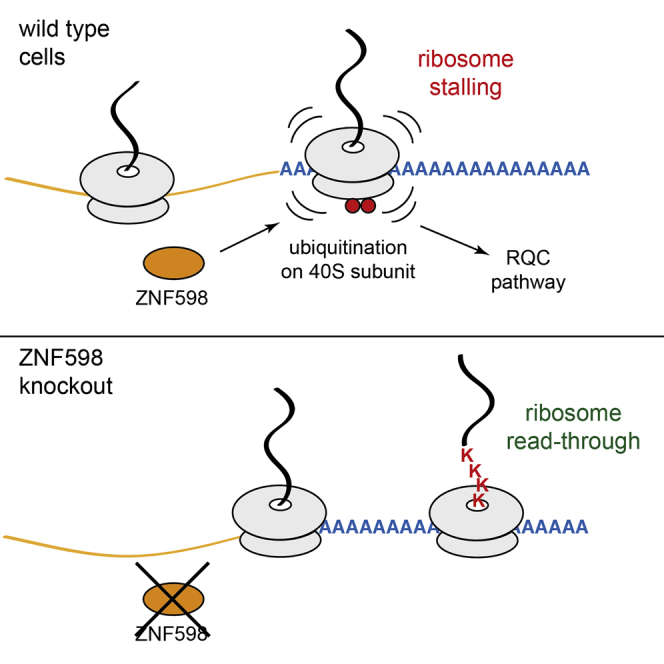
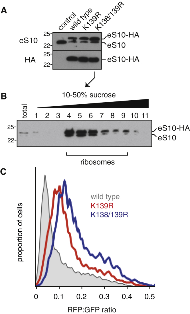
Diverse cellular stressors have been observed to trigger site-specific ubiquitination on several ribosomal proteins. However, the ubiquitin ligases, biochemical consequences, and physiologic pathways linked to these modifications are not known. Here, we show in mammalian cells that the ubiquitin ligase ZNF598 is required for ribosomes to terminally stall during translation of poly(A) sequences. ZNF598-mediated stalling initiated the ribosome-associated quality control (RQC) pathway for degradation of nascent truncated proteins. Biochemical ubiquitination reactions identified two sites of mono-ubiquitination on the 40S protein eS10 as the primary ribosomal target of ZNF598. Cells lacking ZNF598 activity or containing ubiquitination-resistant eS10 ribosomes failed to stall efficiently on poly(A) sequences. In the absence of stalling, read-through of poly(A) produces a poly-lysine tag, which might alter the localization and solubility of the associated protein. Thus, ribosome ubiquitination can modulate translation elongation and impacts co-translational quality control to minimize production of aberrant proteins.

Keywords: nonstop mRNA; poly(A); protein quality control; ribosome stalling; translation; ubiquitination.


Figures

None
Graphical abstract
Figure 1
Figure 1
Assay of Terminal Ribosome Stalling at Single-Cell Resolution (A) Diagram of the reporter construct and expected protein products in the absence or presence of terminal stalling. (B) Median RFP:GFP ratio of 20,000 transfected cells transiently expressing the reporter construct containing the indicated test sequences. Error bars represent 68% of the events around the median. (C) Isogenic stable cell lines expressing the (K)0 (in gray) or (KAAA)21 (in blue) reporter for 24 hr were analyzed by flow cytometry. A scatterplot of individual cells (top) and a histogram of GFP:RFP ratio (bottom) are shown. This and all other scatterplots are shown on a bi-exponential scale to better visualize data across the wide range of expression levels seen in these experiments. (D) Immunoblot of (K)0 and (KAAA)21 cells without and with induction of reporter expression with doxycycline for 24 hr. (E) (KAAA)21 expressing cells were treated with 20 μM MG132 for 4 hr and analyzed by immunoblotting for the indicated products. (F) (KAAA)21 cells were subjected to small interfering RNA (siRNA) treatment against the indicated targets for 72 hr, reporter expression induced for 24 hr, and analyzed by immunoblotting for the indicated proteins. See also Figure S1.
Figure 2
Figure 2
ZNF598 Is Required to Initiate the RQC Pathway during Poly(A) Translation (A) (KAAA)21 cells were transfected with control (blue) or ZNF598 (red) siRNAs for 72 hr, reporter expression induced for 24 hr, and analyzed by flow cytometry (left and middle panels) or immunoblotting for the indicated proteins (right panel). (K)0 cells (gray) served as a reference. (B) (KAAA)21 cells were subjected to the indicated siRNA treatment for 72 hr, reporter expression induced for 24 hr, and analyzed by immunoblotting for the indicated proteins. (C) (KAAA)21 reporter expression was induced for 24 hr in wild-type (WT, in blue) and ZNF598 knockout (KO, in red) cells and analyzed by flow cytometry. (D) (KAAA)21 cells knocked out for ZNF598 were transiently transfected with the indicated plasmids and the transfected cells (as judged by a co-transfected BFP construct) were analyzed by flow cytometry after reporter induction for 24 hr. The majority of cells displayed restoration of terminal ribosomal stalling to wild-type levels (circled). See also Figure S2.
Figure 3
Figure 3
ZNF598 Ubiquitinates eS10 In Vitro and In Vivo (A) In vitro ubiquitination reactions of ribosomes using HA-tagged ubiquitin and the indicated factors were analyzed by immunoblotting with anti-HA to detect all new ubiquitin conjugates. The last reaction was also separated on a 10%–50% sucrose gradient and analyzed by immunoblotting for the indicated antigens (right panel). Asterisks indicate primary ribosomal ubiquitin conjugates. (B) Ribosome ubiquitination reactions with His-tagged ubiquitin in the presence (+) or absence (−) of ZNF598 were fractionated to purify ubiquitinated core ribosomal proteins, treated or untreated with the catalytic domain (CD) of the deubiquitinase Usp2, and analyzed by SDS-PAGE and Coomassie blue staining. The major ZNF598-dependent products were identified by mass spectrometry. (C) Ribosomes were ubiquitinated with increasing concentrations of ZNF598 with either HA-tagged ubiquitin or methyl-ubiquitin, and analyzed by immunoblotting for the indicated ribosomal proteins. (D) eS10 ubiquitination status in WT and ZNF598 KO cells was analyzed by immunoblotting. Two exposures are shown. (E) WT or ZNF598 KO cells were transfected with HA-ubiquitin and treated with nothing, 100 μg/ml cycloheximide, or 1 mM DTT for 2 hr. Anti-eS10 immunoblots are shown for total cell lysate and anti-HA affinity purified products. The positions of ubiquitin- or HA-ubiquitin-modified eS10 are indicated. See also Figure S3.
Figure 4
Figure 4
Ubiquitination of eS10 Facilitates Initiation of the RQC Pathway (A) Cytosol from cells stably expressing different HA-tagged variants of eS10 was analyzed by immunoblotting for eS10 and the HA tag. (B) Cytosol from cells stably expressing the HA-tagged K138/139R mutant of eS10 was separated on a 10%–50% sucrose gradient and immunoblotted for eS10. The fractions containing ribosomes are indicated. Similar results were seen for other eS10 variants (not shown). (C) The indicated HA-eS10 expressing cells were transfected with the (KAAA)20 reporter and analyzed by flow cytometry 24 hr later. The RFP:GFP ratio of all transfected cells is shown as a histogram (eS10-HA in gray, eS10-K139R-HA in red, eS10-K138/139R-HA in blue). See also Figure S4.

Similar articles

Cited by

References

    1. Arthur L., Pavlovic-Djuranovic S., Smith-Koutmou K., Green R., Szczesny P., Djuranovic S. Translational control by lysine-encoding A-rich sequences. Sci. Adv. 2015;1:e1500154. - PMC - PubMed
    1. Bengtson M.H., Joazeiro C.A. Role of a ribosome-associated E3 ubiquitin ligase in protein quality control. Nature. 2010;467:470–473. - PMC - PubMed
    1. Brandman O., Hegde R.S. Ribosome-associated protein quality control. Nat. Struct. Mol. Biol. 2016;23:7–15. - PMC - PubMed
    1. Brandman O., Stewart-Ornstein J., Wong D., Larson A., Williams C.C., Li G.-W., Zhou S., King D., Shen P.S., Weibezahn J. A ribosome-bound quality control complex triggers degradation of nascent peptides and signals translation stress. Cell. 2012;151:1042–1054. - PMC - PubMed
    1. Chakrabarti O., Hegde R.S. Functional depletion of mahogunin by cytosolically exposed prion protein contributes to neurodegeneration. Cell. 2009;137:1136–1147. - PMC - PubMed