. 2017 Jan 18;93(2):331-347.
doi: 10.1016/j.neuron.2016.12.016. Epub 2017 Jan 5.

Translation of Expanded CGG Repeats into FMRpolyG Is Pathogenic and May Contribute to Fragile X Tremor Ataxia Syndrome

Affiliations

Translation of Expanded CGG Repeats into FMRpolyG Is Pathogenic and May Contribute to Fragile X Tremor Ataxia Syndrome

Chantal Sellier et al. Neuron. .

Abstract

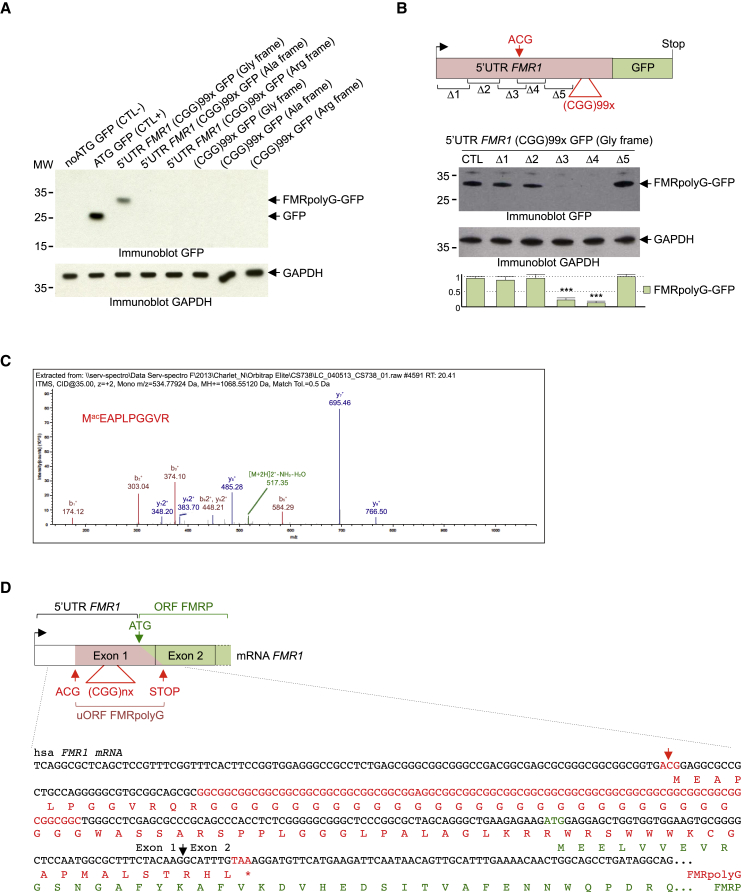
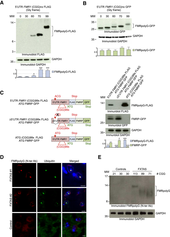
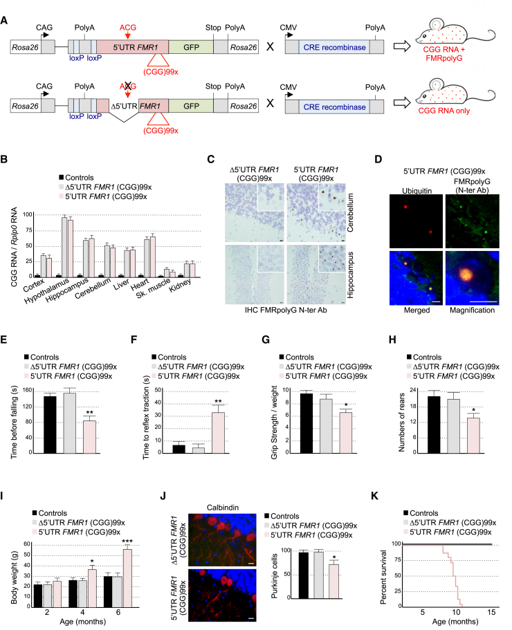
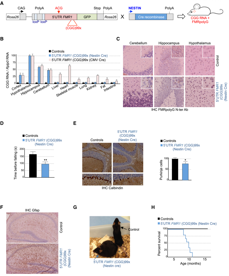
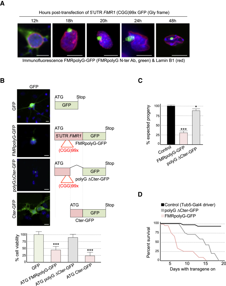
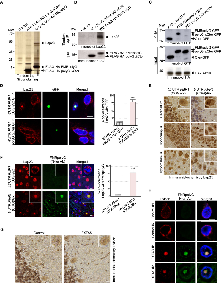
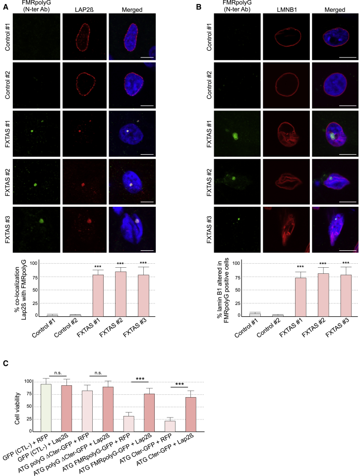
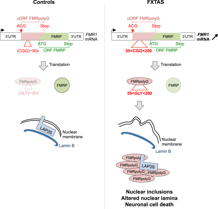
Fragile X-associated tremor/ataxia syndrome (FXTAS) is a neurodegenerative disorder caused by a limited expansion of CGG repeats in the 5' UTR of FMR1. Two mechanisms are proposed to cause FXTAS: RNA gain-of-function, where CGG RNA sequesters specific proteins, and translation of CGG repeats into a polyglycine-containing protein, FMRpolyG. Here we developed transgenic mice expressing CGG repeat RNA with or without FMRpolyG. Expression of FMRpolyG is pathogenic, while the sole expression of CGG RNA is not. FMRpolyG interacts with the nuclear lamina protein LAP2β and disorganizes the nuclear lamina architecture in neurons differentiated from FXTAS iPS cells. Finally, expression of LAP2β rescues neuronal death induced by FMRpolyG. Overall, these results suggest that translation of expanded CGG repeats into FMRpolyG alters nuclear lamina architecture and drives pathogenesis in FXTAS.

Keywords: RAN translation; microsatellite expansion; near-cognate codon; neurodegeneration.


Figures

Figure 1
Figure 1
Translation of CGG Repeats Initiates at an Upstream Near-Cognate Codon (A) Immunoblotting against GFP on the soluble lysate fraction of HeLa cells transfected for 24 hr with expanded CGG repeats embedded, or not, in the 5′ UTR of FMR1 and fused in all three possible frames with the GFP deleted of its ATG. (B) Upper, schemes of the FMR1 5′ UTR deletion constructs tested. Middle, immunoblotting against GFP on the soluble lysate fraction of HeLa cells transfected for 24 hr with mutants of FMR1 5′ UTR containing expanded CGG repeats fused to the GFP in the glycine frame. Lower, quantification of FMRpolyG-GFP expression reported to GAPDH. (C) LC-MS/MS spectra of the N-terminal part of the immunoprecipitated and trypsin-digested protein translated from expanded CGG embedded in the 5′ UTR of FMR1. (D) Scheme and partial nucleotide sequence of human FMR1 exons 1 and 2. Amino acid sequence of FMR1 uORF translated into FMRpolyG is indicated in red. Amino acid sequence of the beginning of the FMRP ORF is indicated in green. Error bars indicate SEM of three independent transfections. Student’s t test, ∗∗∗ indicates p < 0.001.
Figure 2
Figure 2
A Minimum of 60 Expanded CGG Repeats Is Required to Detect FMRpolyG (A) Upper, immunoblotting against the FLAG tag on the soluble lysate fraction of HeLa cells transfected for 24 hr with various lengths of expanded CGG repeats embedded in the 5′ UTR of FMR1 and fused in the glycine frame with the FLAG tag. Middle, control immunoblotting against GAPDH. Lower, quantification of FMRpolyG-FLAG levels reported to GAPDH. (B) Identical to (A), but with CGG repeats embedded in the 5′ UTR of FMR1 fused in the glycine frame with a GFP tag instead of a FLAG tag. (C) Left, schemes of the 5′ UTR of FMR1 constructs with ACG mutations. FMRpolyG uORF is FLAG-tagged in the glycine frame, while FMRP ORF is GFP-tagged. Right, upper, and middle, immunoblotting against FLAG, GFP, and GAPDH on the soluble lysate fraction of HeLa cells transfected for 24 hr with mutants of the 5′ UTR of FMR1; expanded CGG repeats are fused to the FLAG tag in the glycine frame, while the downstream FMRP ORF is fused to the GFP. Right lower, quantification of FMRpolyG-FLAG and FMRP-GFP levels reported to GAPDH. (D) Immunofluorescence against the FMRpolyG N terminus and ubiquitin on brain sections (hippocampal area) of FXTAS or control individuals. Scale bars, 10 μm. Nuclei were counter-stained with DAPI. (E) Immunoblotting against the FMRpolyG N terminus of insoluble fraction of brain lysate (cerebellum area) of FXTAS and age-matched individuals. The numbers of expanded CGG in FMR1 are indicated as # CGG. Error bars indicate SEM of three independent transfections. Student’s t test, indicates p < 0.05, ∗∗∗ indicates p < 0.001.
Figure 3
Figure 3
Expression of FMRpolyG Is Pathogenic in Mice (A) Schemes of the mouse transgene constructs. (B) Quantitative RT-PCR analysis of transgene expression relative to the RplpO mRNA in different brain areas and tissues of 6-month-old control (n = 3), bigenic CMV-cre/full-length (n = 3), or mutant (n = 3) FMR1 5′ UTR transgenic mice. (C) Immunohistochemistry against FMRpolyG N terminus of cerebellum and hippocampus areas of 6-month-old bigenic CMV-cre/full-length or mutant FMR1 5′ UTR transgenic mice. Scale bars, 10 μm. Sections were counter-stained with Nissl staining. (D) Immunofluorescence against FMRpolyG N terminus and ubiquitin on cerebellum areas of 6-month-old bigenic CMV-cre/full-length or mutant FMR1 5′ UTR transgenic mice. Scale bars, 10 μm. Nuclei were counter-stained with DAPI. (E) Rotarod test: the time before falling from a rotating rod of 3-month-old control (n = 8), bigenic CMV-cre/full-length (n = 9), or mutant (n = 9) FMR1 5′ UTR transgenic male mice. (F) String test: the time to gain hindlimb traction for forelimb-hanging 3-month-old control (n = 8), bigenic CMV-cre/full-length (n = 9), or mutant (n = 9) FMR1 5′ UTR transgenic male mice. (G) Grip test: the maximal force relative to mouse body weight exerted to release 3-month-old control (n = 8), bigenic CMV-cre/full-length (n = 9), or mutant (n = 9) FMR1 5′ UTR transgenic male mice holding a grid with their forepaws. (H) Open field: number of rears during 5 min observation in open field of 3-month-old control (n = 8), bigenic CMV-cre/full-length (n = 9), or mutant (n = 9) FMR1 5′ UTR transgenic male mice. (I) Body weight of 2-, 4-, and 6-month-old control (n = 6), bigenic CMV-cre/full-length (n = 6), or mutant (n = 6) FMR1 5′ UTR transgenic male mice. (J) Left, immunofluorescence labeling of calbindin of cerebellum sections of 9-month-old bigenic CMV-cre/full-length or mutant FMR1 5′ UTR transgenic mice. Scale bars, 10 μm. Nuclei were counter-stained with DAPI. Right, quantification of Purkinje cells (n = 100) in cerebellum sections of 9-month-old bigenic CMV-cre/full-length (n = 3) or mutant FMR1 5′ UTR (n = 3) transgenic mice. (K) Kaplan-Meier survival curve of control (n = 15), bigenic CMV-cre/full-length (n = 15), or mutant (n = 15) FMR1 5′ UTR male and female transgenic mice. Error bars indicate SEM. Student’s t test, indicates p < 0.05, ∗∗ indicates p < 0.01 and ∗∗∗ indicates p < 0.001.
Figure 4
Figure 4
Neuronal Expression of FMRpolyG Is Pathogenic in Mice (A) Schemes of the mouse transgene constructs. (B) Quantitative RT-PCR analysis of transgene expression relative to the RplpO mRNA in different tissues of 6-month-old control (n = 3), bigenic CMV-cre/full-length FMR1 5′ UTR (n = 3), or bigenic Nestin-cre full-length FMR1 5′ UTR (n = 3) mice. (C) Immunohistochemistry against FMRpolyG N terminus in the cerebellum, hippocampus, and hypothalamus of 6-month-old bigenic Nestin-cre/full-length FMR1 5′ UTR mice. Scale bars, 10 μm. Sections were counter-stained with H&E staining. (D) Rotarod test: time before falling of 3-month-old control (n = 6) or bigenic Nestin-cre/full-length FMR1 5′ UTR (n = 6) male mice. (E) Left, immunohistochemistry against calbindin of cerebellum sections of 10-month-old control or bigenic Nestin-cre/full-length FMR1 5′ UTR mice. Scale bars, 10 μm. Sections were counter-stained with H&E staining. Right, quantification of Purkinje cells (n = 50) in cerebellum sections of 10-month-old control (n = 3) or bigenic Nestin-cre/full-length FMR1 5′ UTR (n = 3) mice. (F) Immunohistochemistry against Gfap of hippocampal sections of 10-month-old control or bigenic Nestin-cre/full-length FMR1 5′ UTR mice. Scale bars, 50 μm. Sections were counter-stained with H&E staining. (G) Representative image of 8-month-old control or bigenic Nestin-cre/full-length FMR1 5′ UTR mice. (H) Kaplan-Meier survival curves of control (n = 10) or bigenic Nestin-cre/full-length FMR1 5′ UTR (n = 10) male and female mice. Error bars indicate SEM. Student’s t test, indicates p < 0.05, ∗∗ indicates p < 0.01.
Figure 5
Figure 5
FMRpolyG Toxicity Is Influenced by Its Carboxyl Terminus (A) Immunofluorescence against FMRpolyG N terminus and Lmnb1 in primary cultures of E18 mouse cortical neurons transfected for the indicated time period with expanded CGG repeats embedded within the 5′ UTR of FMR1 and fused to the GFP in the glycine frame. Scale bars, 10 μm. Nuclei were counter-stained with DAPI. (B) Left, representative images of primary cultures of E18 mouse cortical neurons transfected with GFP or ATG-driven FMRpolyG-GFP full-length or mutants. Scale bars, 10 μm. Nuclei were counter-stained with DAPI. Right, schemes of the mutant constructs of FMRpolyG-GFP. The Nter construct corresponds to the N-terminal part of FMRpolyG including its polyglycine repeats fused to the GFP. The Cter construct corresponds to the last 42 amino acids of FMRpolyG fused to the GFP. Lower, quantification of neuronal cell viability of GFP-positive (n = 100 cells, three independent transfections) transfected E18 mouse cortical neurons. (C) Progeny eclosion ratio (n = 100, three independent crosses) of Drosophila ubiquitously expressing FMRpolyG, either full-length or deleted of its C terminus compared to control driver line (Actin5C-Gal4/+). (D) Kaplan-Meier survival curve of Drosophila expressing FMRpolyG full-length or deleted of its C terminus compared to control driver line (Tub5-Gal4/+). Error bars indicate SEM. Student’s t test, indicates p < 0.05, ∗∗∗ indicates p < 0.001.
Figure 6
Figure 6
FMRpolyG Interacts with LAP2β and Alters Its Nuclear Localization (A) Silver staining of proteins captured through consecutive anti-FLAG and anti-HA affinity purification steps from N2A cells transfected for 24 hr with ATG-driven FLAG-HA-tagged FMRpolyG, either full-length or deleted of its C terminus. (B) Immunoblotting against endogenous Lap2β protein of tandem-tag purified proteins from N2A cells transfected for 24 hr with ATG-driven FLAG-HA-tagged FMRpolyG, either full-length or deleted of its C terminus. (C) Immunoblotting against the HA or GFP tags of HA-tagged immunoprecipitated proteins from the soluble lysate fraction of N2A cells transfected for 24 hr with HA-LAP2β and ATG-driven FMRpolyG-GFP, either full-length or deleted of its N or C terminus. (D) Left, GFP fluorescence and immunofluorescence against endogenous Lap2β in primary cultures of E18 mouse cortical neurons transfected with expanded CGG repeats embedded within the 5′ UTR of FMR1 either full-length or deleted of FMRpolyG C terminus and fused to the GFP in the glycine frame. Nuclei were counterstained with DAPI. Right, quantification of co-localization of Lap2β with GFP inclusions in transfected E18 mouse cortical neurons (n = 100 neurons, three independent transfections). (E) Immunohistochemistry against Lap2β of cerebellum, hippocampal and hypothalamic areas of 9-month-old bigenic CMV-cre/full-length or mutant FMR1 5′ UTR transgenic mice. Sections were counter-stained with H&E staining. (F) Left, immunofluorescence against FMRpolyG N terminus and Lap2β on hippocampal areas of 9-month-old bigenic CMV-cre/full-length or mutant FMR1 5′ UTR transgenic mice. Nuclei were counter-stained with DAPI. Right, quantification of co-localization of Lap2β with FMRpolyG in bigenic CMV-cre/full-length or mutant FMR1 5′ UTR transgenic mice (n = 50 neurons, three mice). (G) Immunohistochemistry against LAP2β of cerebellum areas of FXTAS individual or age-matched control. Sections were counter-stained with H&E staining. (H) Immunofluorescence against FMRpolyG N terminus and LAP2β on brain sections (hippocampal area) of FXTAS patients or age-matched controls. Nuclei were counter-stained with DAPI. Scale bars, 10 μm. Error bars indicate SEM. Student’s t test, ∗∗∗ indicates p < 0.001.
Figure 7
Figure 7
LAP2β Rescues Neuronal Cell Death Induced by FMRpolyG (A) Upper, immunofluorescence against FMRpolyG N terminus and LAP2β on neuronal cultures differentiated 40 days from iPS cells of FXTAs patients or control individuals. Lower, quantification of LAP2β co-localization with FMRpolyG in neurons from iPSC of FXTAS and control individuals (n = 100 neurons, three independent cultures). (B) Upper, immunofluorescence against FMRpolyG N terminus and LMNB1 on neuronal cultures differentiated 40 days from iPS cells of FXTAs patients or control individuals. Lower, quantification of lamin B1 alteration in FMRpolyG-positive cells in neurons from iPSC of FXTAS and control individuals (n = 100 neurons, three independent cultures). (C) Cell viability of neuronal N2A cells transfected (n = 3 transfections) with ATG-driven FMRpolyG-GFP either full-length or deleted of its N or C terminus and with a plasmid expressing RFP as control or Ha-tagged LAP2β. Error bars indicate SEM. Student’s t test, ∗∗∗ indicates p < 0.001.
Figure 8
Figure 8
A Working Model for Pathogenicity in FXTAS Expanded CGG repeats are translated into a polyglycine-containing protein, FMRpolyG, through initiation to a non-canonical ACG codon located upstream of the CGG repeats. In FXTAS, higher expression of FMR1 mRNA and increased translation and stability of the expanded CGG repeats result in accumulation of FMRpolyG in nuclear aggregates that sequester the LAP2β protein and alter the nuclear lamina architecture.

References

    1. Arocena D.G., Iwahashi C.K., Won N., Beilina A., Ludwig A.L., Tassone F., Schwartz P.H., Hagerman P.J. Induction of inclusion formation and disruption of lamin A/C structure by premutation CGG-repeat RNA in human cultured neural cells. Hum. Mol. Genet. 2005;14:3661–3671. - PubMed
    1. Ash P.E., Bieniek K.F., Gendron T.F., Caulfield T., Lin W.L., Dejesus-Hernandez M., van Blitterswijk M.M., Jansen-West K., Paul J.W., 3rd, Rademakers R. Unconventional translation of C9ORF72 GGGGCC expansion generates insoluble polypeptides specific to c9FTD/ALS. Neuron. 2013;77:639–646. - PMC - PubMed
    1. Aspden J.L., Eyre-Walker Y.C., Phillips R.J., Amin U., Mumtaz M.A., Brocard M., Couso J.P. Extensive translation of small Open Reading Frames revealed by Poly-Ribo-Seq. Elife. 2014;3:e03528. - PMC - PubMed
    1. Barbosa C., Peixeiro I., Romão L. Gene expression regulation by upstream open reading frames and human disease. PLoS Genet. 2013;9:e1003529. - PMC - PubMed
    1. Buijsen R.A., Sellier C., Severijnen L.A., Oulad-Abdelghani M., Verhagen R.F., Berman R.F., Charlet-Berguerand N., Willemsen R., Hukema R.K. FMRpolyG-positive inclusions in CNS and non-CNS organs of a fragile X premutation carrier with fragile X-associated tremor/ataxia syndrome. Acta Neuropathol. Commun. 2014;2:162. - PMC - PubMed

Publication types

MeSH terms

Supplementary concepts