

Challenging cardiac function post-spinal cord injury with dobutamine
- PMID: 28065654
- PMCID: PMC5481490
- DOI: 10.1016/j.autneu.2016.12.005
Challenging cardiac function post-spinal cord injury with dobutamine
Abstract
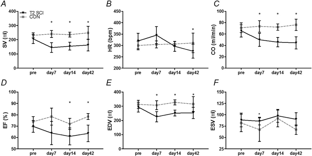
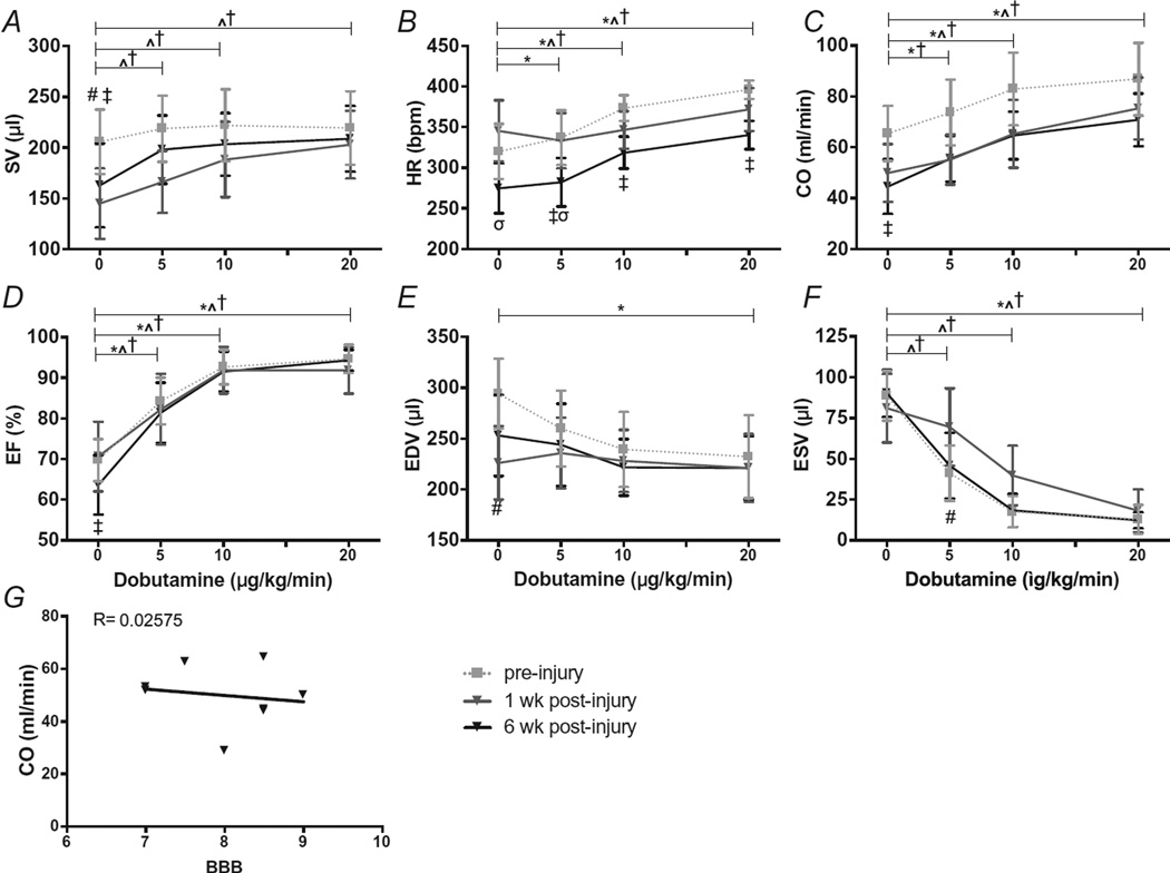
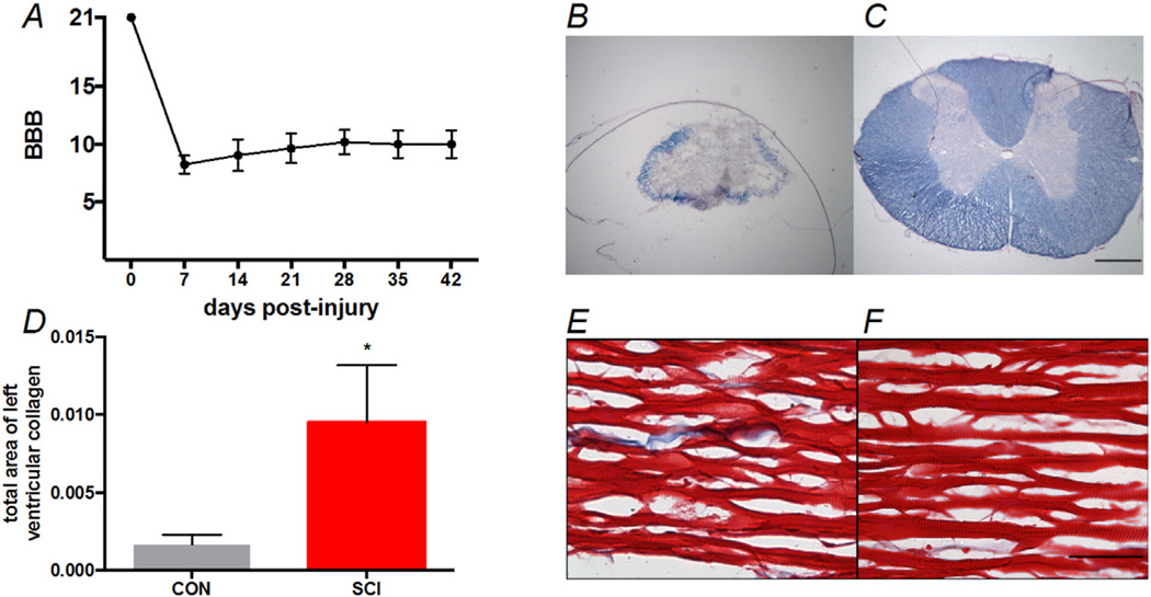
There is general consensus that spinal cord injuries (SCI) above T6 result in altered sympathetic control of the heart, which negatively influences cardiac structure and function. To by-pass disrupted circuitry and investigate cardiac responses under enhanced sympathetic activity we utilized dobutamine (DOB) stress echocardiography. Animals were divided into a T2, 25g-cm contusive SCI (SCI) or an uninjured control (CON) group. Echocardiography was performed pre-SCI and at 1, 2 and 6weeks post-SCI. Increasing doses of DOB (5, 10 & 20μg/min/kg) were infused intravenously pre-SCI and at 1 and 6weeks post-SCI. Parasternal-short axis images were used to compare group differences in systolic function and track changes in response to SCI and DOB over time. One week post-SCI, stroke volume (SV), end diastolic volume (EDV), cardiac output (CO) and ejection fraction (EF) were all reduced compared to CON and these deficits persisted to 6weeks. We also found an increase in collagen deposition at 6weeks post SCI. Pre-SCI, DOB elicited a decrease in EDV and increases in CO, EF and HR but not SV. At 6weeks following SCI, in addition to increases in CO, EF and HR, DOB also induced increases in SV. This is the first report, to our knowledge, of DOB responses in a contusive SCI model with persistent cardiac impairments. The return of CO to pre-SCI levels and the substantial increase in SV at low DOB dosages shows that impaired descending control of the heart is directly contributing to reduced resting SV after SCI.
Keywords: Cardiac; Echocardiography; Spinal cord injury.
Copyright © 2016 Elsevier B.V. All rights reserved.
Conflict of interest statement
No conflicts of interest, financial or otherwise, are declared by the author(s).
Figures
Similar articles
-
A comparison of passive hindlimb cycling and active upper-limb exercise provides new insights into systolic dysfunction after spinal cord injury.Am J Physiol Heart Circ Physiol. 2017 Nov 1;313(5):H861-H870. doi: 10.1152/ajpheart.00046.2017. Epub 2017 Jul 14. Am J Physiol Heart Circ Physiol. 2017. PMID: 28710067 Free PMC article.
-
Markers of susceptibility to cardiac arrhythmia in experimental spinal cord injury and the impact of sympathetic stimulation and exercise training.Auton Neurosci. 2021 Nov;235:102867. doi: 10.1016/j.autneu.2021.102867. Epub 2021 Aug 10. Auton Neurosci. 2021. PMID: 34399294
-
Micro-CT imaging assessment of dobutamine-induced cardiac stress in rats.J Pharmacol Toxicol Methods. 2011 Jan-Feb;63(1):24-9. doi: 10.1016/j.vascn.2010.04.002. Epub 2010 Apr 23. J Pharmacol Toxicol Methods. 2011. PMID: 20399875 Free PMC article.
-
Vitamin C restores the contractile response to dobutamine and improves myocardial efficiency in patients with heart failure after anterior myocardial infarction.Am Heart J. 2007 Oct;154(4):645.e1-8. doi: 10.1016/j.ahj.2007.07.005. Am Heart J. 2007. PMID: 17892985
-
Elevated vascular resistance and afterload reduce the cardiac output response to dobutamine in early growth-restricted rats in adulthood.Br J Nutr. 2011 Nov;106(9):1374-82. doi: 10.1017/S0007114511001784. Epub 2011 Jun 1. Br J Nutr. 2011. PMID: 21736800
Cited by
-
Chronic, complete cervical6-7 cord transection: distinct autonomic and cardiac deficits.J Appl Physiol (1985). 2018 Jun 1;124(6):1471-1482. doi: 10.1152/japplphysiol.01104.2017. Epub 2018 Feb 22. J Appl Physiol (1985). 2018. PMID: 29470149 Free PMC article.
-
Temporal analysis of cardiovascular control and function following incomplete T3 and T10 spinal cord injury in rodents.Physiol Rep. 2018 Mar;6(6):e13634. doi: 10.14814/phy2.13634. Physiol Rep. 2018. PMID: 29595874 Free PMC article.
-
A comparison of passive hindlimb cycling and active upper-limb exercise provides new insights into systolic dysfunction after spinal cord injury.Am J Physiol Heart Circ Physiol. 2017 Nov 1;313(5):H861-H870. doi: 10.1152/ajpheart.00046.2017. Epub 2017 Jul 14. Am J Physiol Heart Circ Physiol. 2017. PMID: 28710067 Free PMC article.
-
Development of a Spinal Cord Injury Model Permissive to Study the Cardiovascular Effects of Rehabilitation Approaches Designed to Induce Neuroplasticity.Biology (Basel). 2021 Oct 7;10(10):1006. doi: 10.3390/biology10101006. Biology (Basel). 2021. PMID: 34681105 Free PMC article.
-
The susceptibility of cardiac arrhythmias after spinal cord crush injury in rats.Exp Neurol. 2022 Nov;357:114200. doi: 10.1016/j.expneurol.2022.114200. Epub 2022 Aug 8. Exp Neurol. 2022. PMID: 35952765 Free PMC article.
References
-
- Basso DM, Beattie MS, Bresnahan JC. A sensitive and reliable locomotor rating scale for open field testing in rats. J. Neurotrauma. 1995;12:1–21. - PubMed
-
- Conrad CH, Brooks WW, Hayes JA, Sen S, Robinson KG, Bing OH. Myocardial fibrosis and stiffness with hypertrophy and heart failure in the spontaneously hypertensive rat. Circulation. 1995;91:161–170. - PubMed
-
- Dampney RA. Functional organization of central pathways regulating the cardiovascular system. Physiol. Rev. 1994;74:323–364. - PubMed
Publication types
MeSH terms
Substances
Grants and funding
LinkOut - more resources
Full Text Sources
Other Literature Sources
Medical
