. 2017 May;13(4):1495-1506.
doi: 10.1016/j.nano.2016.12.019. Epub 2017 Jan 5.

Mechanisms that determine nanocarrier targeting to healthy versus inflamed lung regions

Affiliations

Mechanisms that determine nanocarrier targeting to healthy versus inflamed lung regions

Jacob S Brenner et al. Nanomedicine. 2017 May.

Abstract

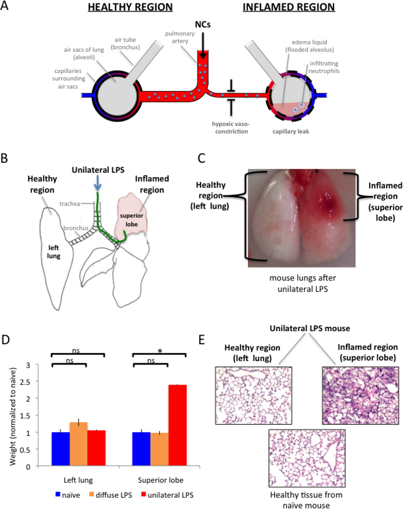
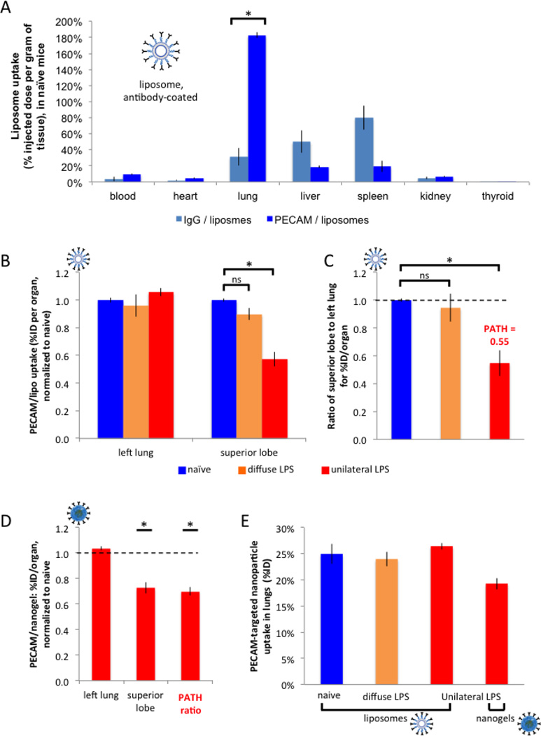
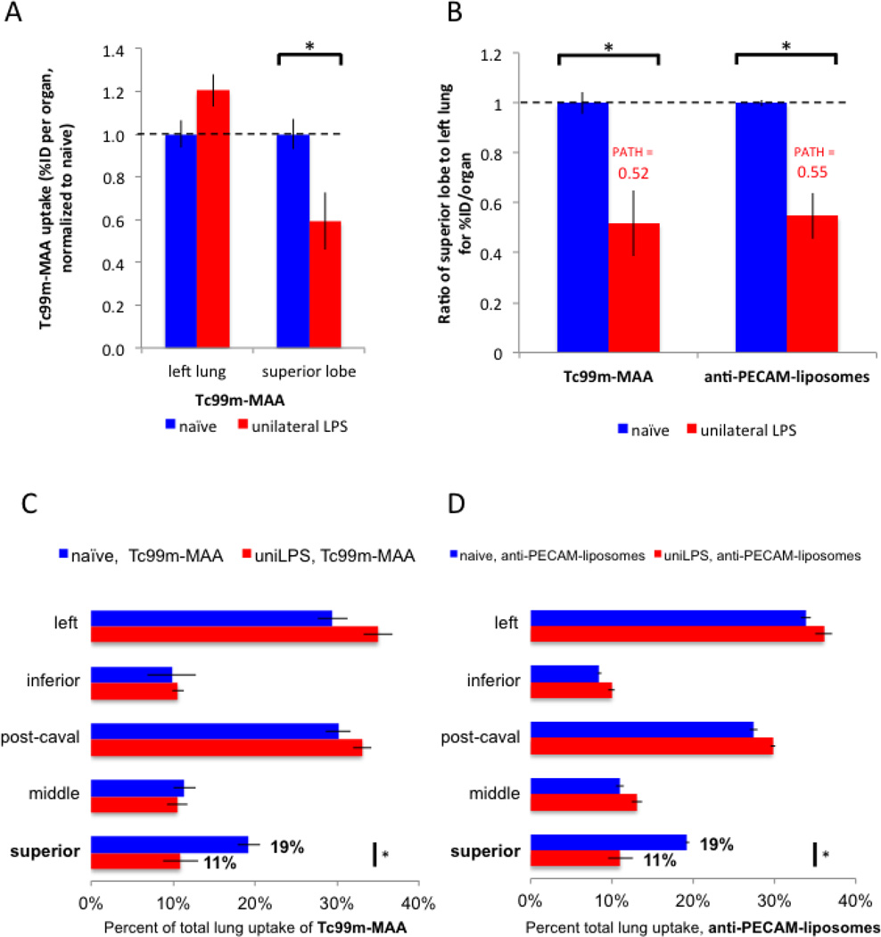
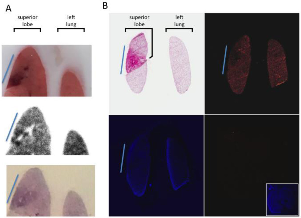
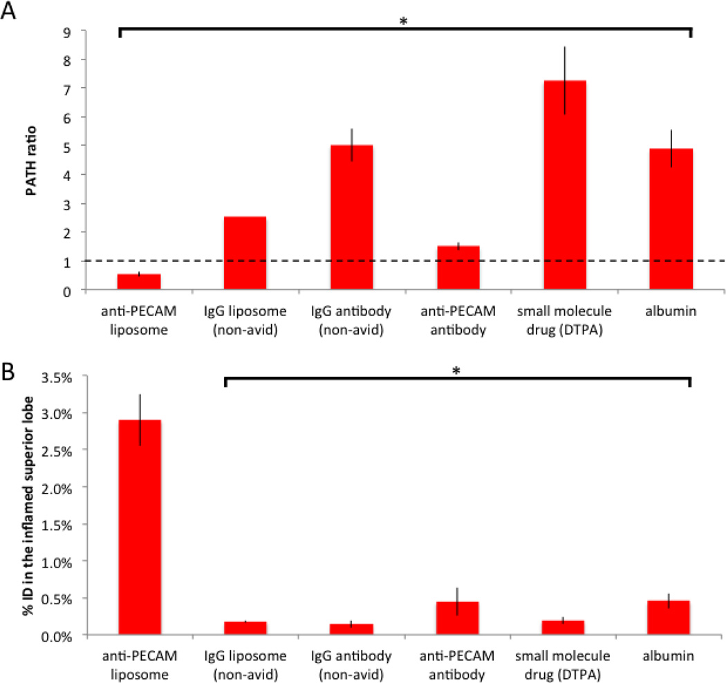
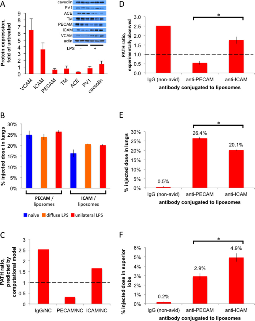
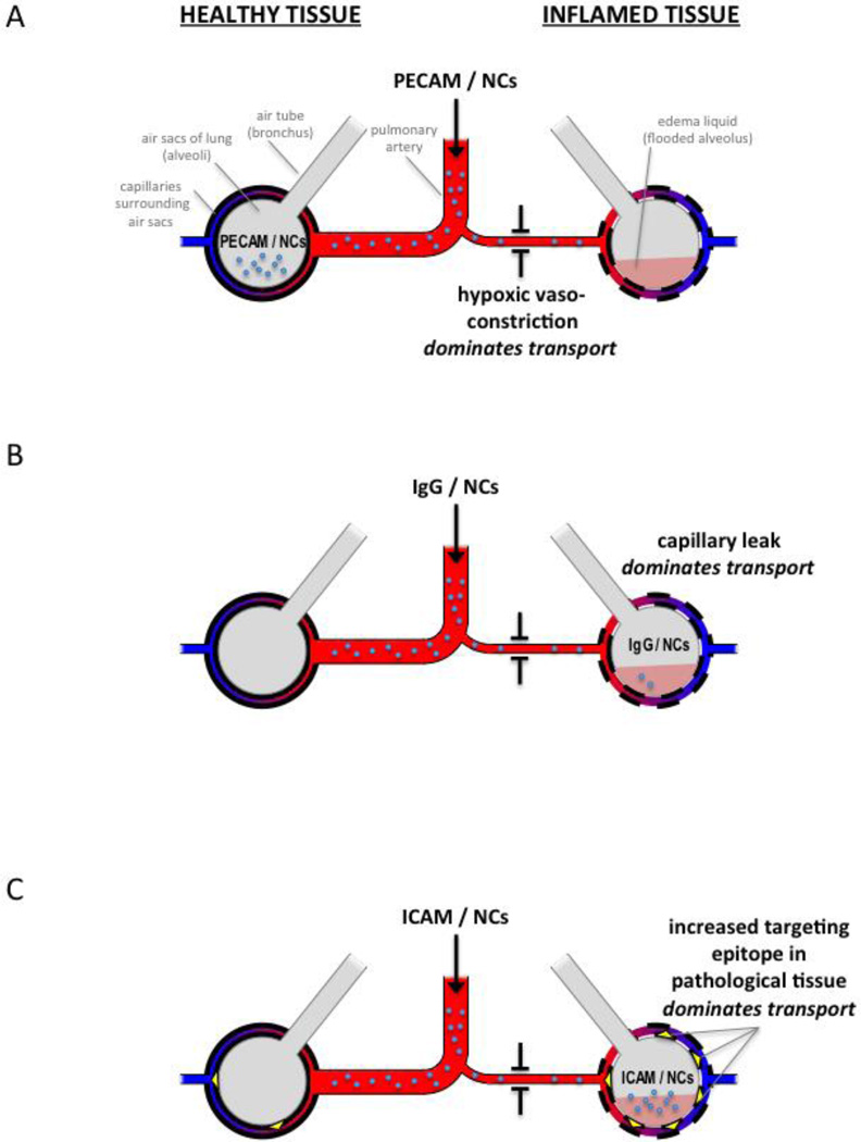
Inflamed organs display marked spatial heterogeneity of inflammation, with patches of inflamed tissue adjacent to healthy tissue. To investigate how nanocarriers (NCs) distribute between such patches, we created a mouse model that recapitulates the spatial heterogeneity of the inflammatory lung disease ARDS. NCs targeting the epitope PECAM strongly accumulated in the lungs, but were shunted away from inflamed lung regions due to hypoxic vasoconstriction (HVC). In contrast, ICAM-targeted NCs, which had lower whole-lung uptake than PECAM/NCs in inflamed lungs, displayed markedly higher NC levels in inflamed regions than PECAM/NCs, due to increased regional ICAM. Regional HVC, epitope expression, and capillary leak were sufficient to predict intra-organ of distribution of NCs, antibodies, and drugs. Importantly, these effects were not observable with traditional spatially-uniform models of ARDS, nor when examining only whole-organ uptake. This study underscores how examining NCs' intra-organ distribution in spatially heterogeneous animal models can guide rational NC design.

Keywords: ARDS; Inflammation; Nano-bio interface; Nanocarriers; Nanoparticle biological interactions; Nanoparticles; Patchy; Spatial heterogeneity; Whole organ distribution.


Figures

Figure 1
Figure 1. A new mouse model of ARDS for testing whether NCs preferentially distribute to inflamed or healthy lung regions
(A) Schematic of lungs in ARDS, with both healthy regions of lung tissue (left side) and inflamed regions (right side) in the same individual. IV-injected NCs reach the lung via the pulmonary artery (center), which has branches leading to each lung region. Lung regions are depicted as air sacs (large grey circles) surrounded by capillaries (tubes with red-to-blue gradient). The pathological region experiences hypoxic vasoconstriction (arteriolar narrowing), capillary leak (increased permeability of the capillaries), and filling of the air sacs with edema liquid (pink) and neutrophils. (B) Diagram of unilateral LPS instillation, dorsal view. A custom 0.024” OD catheter (green) is threaded through the mouth and into the superior lobe, whereupon LPS is instilled. (C) Mouse lungs (dorsal view) that received unilateral LPS display a healthy region that is indistinguishable from naive (left lung; white), and a severely inflamed region (superior lobe; red). (D) To quantify the ARDS phenotype of edema liquid, we measured the weights of each lung region. Compared to naive (blue bars), the unilateral LPS model (red bars) displays no increased weight in the healthy region (left lung), but a 2.4-fold increase in weight of the inflamed region (superior lobe), due to edema liquid filling the air sacs of the inflamed region. The classical model of “diffuse LPS” (which is spatially homogeneous) (orange bars) displays no change in weight. (E) To observe the ARDS phenotype of neutrophilic infiltration, unilateral LPS mouse lungs were fixed and H&E-stained, which displays neutrophils as round purple cells. The healthy lung tissue (left lung) of these mice displays no neutrophilic infiltrate (left panel) when compared to a naive mouse (bottom panel), while the inflamed tissue (superior lobe) has every air sac filled with neutrophils (right panel). *, p < 0.01. ns, non-significant.
Figure 2
Figure 2. PECAM/NCs markedly concentrate in the lungs, but preferentially accumulate in healthy rather than inflamed lung tissue
(A) (A) Healthy (naïve) mice were IV-injected with liposomes (inset; labeled with I125) conjugated to anti-PECAM antibodies or control antibodies, sacrificed 30 minutes later, and organ uptake was measured by gamma counter. Anti-PECAM liposomes display 5.9-fold higher lung concentratinos than control liposomes. (B) 24 hours after either unilateral LPS (red bars), diffuse LPS (orange), or sham (naive; blue), mice were IV-injected with anti-PECAM-liposomes, sacrificed 30 minutes later, and then lung lobes were measured as in A. For each mouse, we calculated the ratio of superior lobe to left lung for the percent injected dose (%ID) per organ (“organ” here being a lung lobe), and normalized to naive. (C) For each mouse, we calculated the ratio of superior lobe to left lung for the %ID/organ values in B, normalized to the value for naïve mice. Note that this ratio for unilateral LPS mice (third bar) is defined as the PATH ratio (Pathologically Altered To Healthy ratio), which is the %ID/organ of the pathological (inflamed) tissue divided by the %ID/organ of the healthy tissue, normalized to naive. A PATH ratio < 1 (here it is 0.55), indicates that a NC preferentially accumulates in healthy tissue, while a PATH ratio > 1 implies preferential accumulation in pathological (inflamed) tissue. (D) With the same protocol as in B and C, unilateral LPS mice were injected with an alternative nanocarrier, nanogels, also coated with anti-PECAM antibodies, producing a similar PATH ratio < 1. Only values with unilateral LPS are displayed (red bars). (F) Replotting A-D as %ID shows no differences in whole-lung uptake of PECAM/NCs, regardless of the presence of LPS instillation. *, p < 0.05. ns, non-significant.
Figure 3
Figure 3. PECAM/NCs preferentially accumulate in the healthy lung regions because of hypoxic vasoconstriction in the inflamed regions
(A) Regional blood flow was measured by IV-injecting Tc99m-MAA. The inflamed superior lobe of unilateral LPS mice displays reduced blood flow (lower Tc99m-MAA / organ), likely to due hypoxic vasoconstriction. (B) Replotting the data of A as the ratio of superior lobe to left lung for %ID/organ (first two bars) and comparing this data to the same ratios obtained for PECAM/NCs (third and fourth bars). Note the two PATH ratios are nearly identical. (C and D) To display the correlation of blood flow and PECAM/NC distribution, all 5 lobes from the mice in A are displayed as % of total lung uptake. Mice injected with either Tc99m-MAA (C) or PECAM/NCs (D) display nearly identical patterns, particularly in the superior and left lobes. *, p < 0.05.
Figure 4
Figure 4. Within the inflamed lung lobe, sub-regions with the most severe inflammation receive the least PECAM/NCs
(A) At 24 hours after unilateral LPS instillation, mice were injected with In111-labeled anti-PECAM-liposomes, and 30 minutes later lungs were removed and fresh-frozen. The top panel shows a slice of the tissue block, displaying a very clear sub-lobar inflammatory injury (blue line). DAR (middle panel) of the same slice shows the highly inflamed sub-region has portions with nearly no nanoparticle uptake (white holes) surrounded by other areas with very strong nanoparticle uptake (black). Bottom panel, H&E staining reveals intense neutrophilic infiltrates (dark purple) in this sub-region. (B) Similar protocol to A, but with rhodamine-labeled liposomes. Top left, H&E shows a sub-region of the superior lobe with severe inflammation (blue line). Top right, rhodamine signal the severely pathological sub-region (blue line) has less liposome uptake (less red signal) than the rest of the lobe. Bottom left, DAPI image. Bottom right, Image from an uninjected mouse displays nearly no signal with the same microscope settings as the top right panel, but normal DAPI signal (inset).
Figure 5
Figure 5. IgG/NCs, small molecule drugs, and proteins preferentially accumulate in the most inflamed tissue because of capillary leak
(A) The PATH ratio for unilateral LPS mice for a variety of pharmacological agents shows only PECAM/NCs have a PATH ratio < 1 (shunting away from the most inflamed lung tissue), while all others have PATH ratio values >1 (preferential accumulation in the most inflamed tissue). Note that radiolabeled albumin (sixth bar in this plot) is the gold-standard for measuring capillary leak, and its PATH ratio of 5 here fits with reported capillary leak metrics in severe ARDS. (B) Despite the high PATH ratios, these other agents have markedly lower total mass accumulating in the most inflamed region (superior lobe) than PECAM/NCs, due to the much higher total lung uptake of PECAM/NCs. *, p < 0.01. ns, non-significant. * indicates statistical comparison to naïve control.
Figure 6
Figure 6. ICAM/NCs accumulate preferentially in inflamed lung regions because of increased epitope levels in that region
(A) A screen to identify targeting epitopes that are increased in the lungs of mice after LPS stimulation. Inset, Western blot of whole lung homogenates from either naive mice or mice given IV LPS 24 hours before sacrifice. Each lane represents an individual mouse, 3 mice per condition. The bar graph shows quantification of the inset Western, normalized to actin, and then mean of LPS mice was divided by the mean of naive mice. (B) ICAM/liposomes display increased (1.26x) whole-lung uptake in diffusely inflamed lungs, but still have lower (by 15%) whole-lung uptake than PECAM/liposomes. 24 hours after either unilateral LPS (red bars), diffuse LPS (orange), or sham (naive; blue), mice were IV-injected with I125-labeled liposomes and whole-lung uptake measured 30 minutes later. (C) A multi-compartment pharmacokinetic model (see Supplemental Materials), incorporating our measurements of hypoxic vasoconstriction, capillary leak, and epitope expression, predicts a PATH ratio < 1 for PECAM/NCs (first red bar) but a PATH ratio ∼1 for ICAM/NCs. (D) Experimentally determined PATH ratios show that PECAM/NCs preferentially accumulate in healthy lung tissue (PATH ratio < 1), while ICAM/NCs are preferentially taken up into the inflamed superior lobe (PATH ratio > 1). (E) Whole-lung uptake, shows PECAM/NCs have the greatest total lung uptake. (F) Uptake in the just the intended target lobe, the severely inflamed superior lobe, with ICAM/NCs performing the best. *, p < 0.05.
Figure 7
Figure 7. Three transport mechanisms determine relative distribution of NCs between healthy and inflamed lung regions
Inflamed tissue displays 3 changes that affect NC transport: hypoxic vasoconstriction (depicted as a narrow arteriole), capillary leak (depicted as dashed line of capillary walls), and increased ICAM epitope (yellow triangles in C). (A) PECAM/NCs (small blue circles) bind very rapidly to the capillary endothelium, and thus their distribution is determined by relative blood flow to each lung region. Therefore, PECAM/NC distribution is dominated by hypoxic vasoconstriction in inflamed lung tissue, resulting in preferential accumulation of PECAM/NCs in healthy tissue (depicted, for simplicity, as NCs in the air sac), which implies a PATH ratio < 1. (B) IgG/NCs only accumulate in the lung where there is capillary leak, so capillary leak dominates distribution, resulting in IgG/NCs accumulating in the inflamed tissue (PATH ratio > 1). Compared to the avid lung binding of PECAM/NCs, the leak-induced lung accumulation of IgG/NCs is very low, depicted as relatively few NCs present in the lungs. (C) ICAM/NCs encounter more accessible ICAM epitopes in inflamed tissue, and this effect dominates over hypoxic vasoconstriction, resulting in preferential accumulation in the inflamed lung tissue (PATH ratio > 1). Additionally, ICAM/NCs benefit from avid lung binding, and thus have high levels of overall lung uptake (depicted as a large number of NCs in the lungs).

References

    1. Uddin MJ, Werfel TA, Crews BC, Gupta MK, Kavanaugh TE, Kingsley PJ, et al. Fluorocoxib A loaded nanoparticles enable targeted visualization of cyclooxygenase-2 in inflammation and cancer. Biomaterials. 2016;92:71–80. - PMC - PubMed
    1. Zhou H-F, Yan H, Hu Y, Springer LE, Yang X, Wickline SA, et al. Fumagillin prodrug nanotherapy suppresses macrophage inflammatory response via endothelial nitric oxide. ACS Nano. 2014;8:7305–7317. - PMC - PubMed
    1. Nguyen MM, Carlini AS, Chien M-P, Sonnenberg S, Luo C, Braden RL, et al. Enzyme-Responsive Nanoparticles for Targeted Accumulation and Prolonged Retention in Heart Tissue after Myocardial Infarction. Adv Mater. 2015;27:5547–5552. - PMC - PubMed
    1. Pan D, Pham CTN, Weilbaecher KN, Tomasson MH, Wickline SA, Lanza GM. Contact-facilitated drug delivery with Sn2 lipase labile prodrugs optimize targeted lipid nanoparticle drug delivery. Wiley Interdiscip Rev Nanomed Nanobiotechnol. 2016;8:85–106. - PMC - PubMed
    1. Kowalski PS, Kuninty PR, Bijlsma KT, Stuart MCA, Leus NGJ, Ruiters MHJ, et al. SAINT-liposome-polycation particles, a new carrier for improved delivery of siRNAs to inflamed endothelial cells. Eur J Pharm Biopharm. 2015;89:40–47. - PubMed

MeSH terms