. 2016:127:282-299.

THE JEREMIAH METZGER LECTURE VARICELLA ZOSTER VIRUS: FROM OUTSIDE TO INSIDE

Affiliations

THE JEREMIAH METZGER LECTURE VARICELLA ZOSTER VIRUS: FROM OUTSIDE TO INSIDE

Anne A Gershon et al. Trans Am Clin Climatol Assoc. 2016.

Abstract

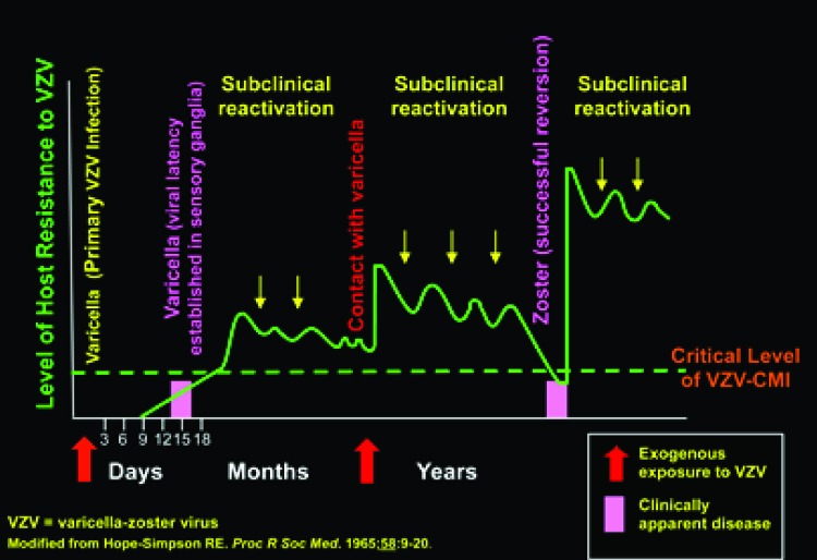
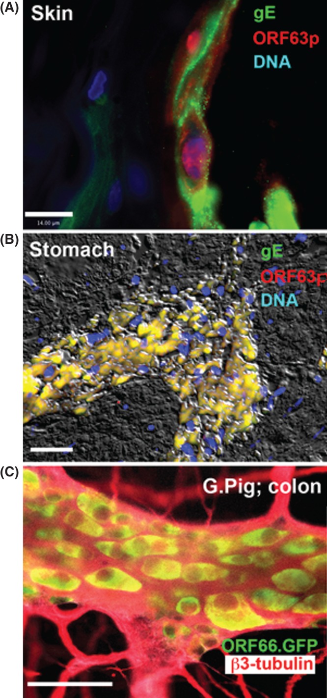
Varicella zoster virus (VZV) gives rise to two diseases, a primary infection, varicella, and a secondary infection, zoster. Morbidity and mortality from VZV in the United States has decreased by 80% to 90% due to the effective use of attenuated live viral vaccines. Because latent VZV continues to reactivate, however, serious VZV-induced disease persists. Newly developed molecular analyses have revealed that zoster is more common than previously realized; moreover, the establishment of VZV latency in neurons, such as those of the enteric nervous system, which do not project to the skin, leads to unexpected, serious, and clandestine manifestations of disease, including perforating gastrointestinal ulcers and intestinal pseudo-obstruction. The development of the first animal model of zoster, in guinea pigs, now enables the pathophysiology of latency and reactivation to be analyzed.


Conflict of interest statement

Potential Conflict of Interest: A.A.G.: Service contracts (molecular VZV diagnosis for vaccine safety): Merck; Ad hoc consulting and chair DSMB: GSK (on VZV). Supported by NIH R01 DK093094.

Figures

Fig. 1
Fig. 1
Transcripts encoding VZV gene products can only be found in segments of gut that contain ganglia. Transcripts and DNA encoding ORF63 were analyzed in surgically removed specimens of colon. ORF63, which is expressed during VZV latency, was analyzed as a marker for the presence of latently infected cells. Transcripts and DNA encoding ORF63 were each detected in a patient with a history of natural varicella (Var), a vaccinated patient (Vac) and in the proximal ganglionated region of bowel from a patient with Hirschsprung’s disease (HSPG). In contrast, neither transcripts nor DNA were detected in the distal aganglionic region of gut of the same patient with HSPG or in the intestine of a newborn infant, which was investigated as a control that neither experienced varicella, nor was vaccinated against it.
Fig. 2
Fig. 2
The hypothesis of Hope-Simpson postulates that internal reactivation of VZV maintains immunity. A person is immunized during an initial attack of varicella but the virus becomes latent in neurons. Periodic subclinical reactivations (contained reversions) keep up immunity. This can be supplemented by external contacts with varicella. Immunity declines, however, as a function of age. This decline allows a periodic reactivation to cross the critical threshold of cell-mediated immunity (CMI) and zoster occurs (successful reversion). The zoster vaccine is designed to boost CMI and deter a periodic reactivation from crossing of the clinical threshold and causing active zoster.
Fig. 3
Fig. 3
Immunoreactivity of VZV proteins in lesions. (A) Human skin. Persistent VZV in the epidermis of a man who died of recurrent episodes of disseminated zoster with granulomas (59). The virus had acquired resistance to acyclovir. The immunoreactivity of a late protein (gE; green) and an immediate early protein (ORF63p; red) can be seen. Note that the infection is lytic and ORF63p is predominantly intranuclear in location. The marker = 14 µm. (B) Human stomach. The fluorescence of VZV was detected in the excised stomach of a 16-year-old vaccinated male. The immunoreactivities of gE (green) and ORF63p (red) are co-localized; the image is merged so that the co-localized fluorescence is yellow. DNA fluorescence is blue. Interference microscopy with white light is superimposed to help with orientation. The marker = 50 µm. (C) Guinea pig colon from an animal in which with VZV-infected lymphocytes were injected intravenously. The VZV expresses green fluorescent protein (GFP) under the control of the ORF66 promoter (courtesy of Dr. Paul Kinchington). b3-tubulin immunoreactivity (red) is used as a neural marker and shows the limits of the ganglia. Only cell bodies have GFP fluorescence (yellow) in the merged image with red fluorescence of b3-tubulin.

References

    1. Gershon A, Breuer J, Cohen JI, et al. Varicella zoster virus infection. Nat Rev Primers. 2015;1:15016–34. - PMC - PubMed
    1. Arvin AM, Moffat JF, Sommer M, et al. Varicella-zoster virus T cell tropism and the pathogenesis of skin infection. Curr Topics Microbiol Immunol. 2010;342:189–209. - PMC - PubMed
    1. Sen N, Mukherjee G, Sen A, et al. Single-cell mass cytometry analysis of human tonsil T cell remodeling by varicella zoster virus. Cell Reports. 2014;8:633–45. - PMC - PubMed
    1. Chen JJ, Zhu Z, Gershon AA, Gershon MD. Mannose 6-phosphate receptor dependence of varicella zoster virus infection in vitro and in the epidermis during varicella and zoster. Cell. 2004;119:915–26. - PubMed
    1. Gershon MD, Gershon AA. VZV infection of keratinocytes: production of cell-free infectious virions in vivo. Curr topics Microbiol Immunol. 2010;342:173–88. - PMC - PubMed

LinkOut - more resources