. 2016 Dec 20:8:310.
doi: 10.3389/fnagi.2016.00310. eCollection 2016.

Early Cognitive/Social Deficits and Late Motor Phenotype in Conditional Wild-Type TDP-43 Transgenic Mice

Affiliations

Early Cognitive/Social Deficits and Late Motor Phenotype in Conditional Wild-Type TDP-43 Transgenic Mice

Julio A Alfieri et al. Front Aging Neurosci. .

Abstract

Frontotemporal Dementia (FTD) and amyotrophic lateral sclerosis (ALS) are two neurodegenerative diseases associated to mislocalization and aggregation of TAR DNA-binding protein 43 (TDP-43). To investigate in depth the behavioral phenotype associated with this proteinopathy, we used as a model transgenic (Tg) mice conditionally overexpressing human wild-type TDP 43 protein (hTDP-43-WT) in forebrain neurons. We previously characterized these mice at the neuropathological level and found progressive neurodegeneration and other features that evoke human TDP-43 proteinopathies of the FTD/ALS spectrum. In the present study we analyzed the behavior of mice at multiple domains, including motor, social and cognitive performance. Our results indicate that young hTDP-43-WT Tg mice (1 month after post-weaning transgene induction) present a normal motor phenotype compared to control littermates, as assessed by accelerated rotarod performance, spontaneous locomotor activity in the open field test and a mild degree of spasticity shown by a clasping phenotype. Analysis of social and cognitive behavior showed a rapid installment of deficits in social interaction, working memory (Y-maze test) and recognition memory (novel object recognition test) in the absence of overt motor abnormalities. To investigate if the motor phenotype worsen with age, we analyzed the behavior of mice after long-term (up to 12 months) transgene induction. Our results reveal a decreased performance on the rotarod test and in the hanging wire test, indicating a motor phenotype that was absent in younger mice. In addition, long-term hTDP-43-WT expression led to hyperlocomotion in the open field test. In sum, these results demonstrate a time-dependent emergence of a motor phenotype in older hTDP-43-WT Tg mice, recapitulating aspects of clinical FTD presentations with motor involvement in human patients, and providing a complementary animal model for studying TDP-43 proteinopathies.

Keywords: TDP-43; amyotrophic lateral sclerosis; animal model; behavior; frontotemporal dementia; proteinopathy; transgenic mice.


Figures
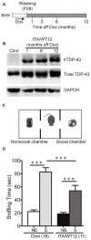
Figure 1
Figure 1
Altered social behavior in TDP-43-WT transgenic (Tg) mice. (A) Experimental design: transgene expression was activated at weaning (postnatal day 28) by removing Dox from water. The behavioral responses of these Tg mice were analyzed at the indicated time points after weaning. (B) Expression of human TAR DNA-binding protein 43 (TDP-43) in Tg mice. Immunoblot of hTDP-43 or total TDP-43 (h+mTDP-43) in cortical RIPA extracts of control (non-Tg) and tTA/WT12 (1, 6 or 12 months off Dox) mice. GAPDH is a loading control. (C) Schematic view of the three-chamber social interaction apparatus, consisting of a black Plexiglas rectangular box with three interconnected chambers. (D) Time spent sniffing the social (S; P21-P28 mouse) or the non–social (NS; black plastic object) stimulus during a 10 min session (test phase) was recorded. 1 month off Dox bigenic mice (tTA/WT12) presented a reduced social interaction time during the session (***p < 0.001, one-way ANOVA/Newman-Keuls post hoc test). Number of animals is indicated in parentheses. The data represent mean ± SEM.
Figure 2
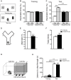
Figure 2
Cognitive impairment in TDP-43-WT Tg mice. (A–C) Novel object recognition test. (A) Scheme of training and test phases for the novel object recognition test. (B) Training day. Both control and TDP-43-WT Tg mice were exposed to two identical objects for 10 min and the time spent exploring each object was recorded. No significant differences in the exploration time (%) of the two objects were found in training phase. (C) Test day. 24 h after training, the recognition memory was measured while the animals were allowed to explore the familiar (Fam) and novel (Nov) objects for 5 min. The exploration time (%) represents the percentage of time that mice spend exploring the object (familiar or novel) respect to the total exploration time (familiar + novel). tTA/WT12 animals displayed a deficit in object recognition memory (***p < 0.001, one-way ANOVA/Newman-Keuls post hoc test). (D–F) Y-maze spontaneous alternation task. (D) Scheme of the Y-maze. (E) Mice were placed at the end of one arm facing the center and allowed to explore the maze freely for 8 min without training, reward or punishment. Entries into each arm were scored and alternation behavior was defined as a complete cycle of consecutive entrances into each of the three arms without repetition. Bigenic tTA/WT12 mice alternated between the arms at the chance (≈50%) level (***p < 0.001 significantly different from control group, Student’s t test; F) Total entries were scored as an index of locomotion activity in the Y maze. (G,H) Step-through inhibitory avoidance test. (G) Scheme of inhibitory avoidance apparatus. (H) During training, each mouse received a footshock (0.2 mA, 50 Hz, 1 s) as it stepped into the dark compartment. Retention test was performed 24 h later. The step-through latency was recorded; footshock was omitted during test session. No significant differences in latency values were found between controls and bigenic animals in either training or test phases, showing intact long term memory for this task (**p < 0.01, ***p < 0.001 Kruskal–Wallis one-way ANOVA followed by individual Mann-Whitney U test). Cognitive behavior was analyzed at 1 month off Dox. Number of animals is indicated in parentheses and data represent mean ± SEM.
Figure 3
Figure 3
Locomotor and exploratory behavior is preserved in young TDP-43-WT mice. To measure general motor function and exploratory activity we used an open field test. Animals were placed in a novel environment during a 20 min session. (A) Total distance traveled, (B) relative center distance and (C) detailed measurement of total distance traveled in time segments of 5 min. No significant differences were found between controls and bigenic animals in locomotion or exploration (p > 0.05, Student’s t test for A,B or repeated-measures ANOVA in C). Number of animals is indicated in parentheses. Data represent mean ± SEM.
Figure 4
Figure 4
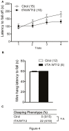
Young TDP-43-WT Tg mice display normal motor coordination, balance and strength. (A) Accelerated rotarod performance (4–40 rpm/5 min). Four trials per test were performed during the test day with a 2 min interval between trials. Latency to fall off the rotarod was recorded. (B) Hanging wire grip test. Grip strength was assessed using a standard wire cage turned upside down. The latency to fall off the wire lid was quantified. A 60 s cutoff time was used. No significant differences were found between control and bigenic animals (p > 0.05, repeated-measures ANOVA in A, Student’s t test in B). (C) Percentage of mice with clasping phenotype reveal mild incidence of spasticity, without reaching significance (Fischer Exact test, p > 0.05); n.s., non-significant differences respect to control group. Number of animals is indicated in parentheses. Data represent mean ± SEM.
Figure 5
Figure 5
TDP-43-WT bigenic mice display normal visual perception and signs of decreased anxiety. (A) Visual perception. Percentage of animals stopping at the edge in the visual cliff test. No significant differences in the response to the edge were found between control and bigenic animals (p > 0.05, Mann-Whitney U test). (B–F) Elevated plus maze test. Mice were placed at the center and allowed to explore the maze freely for 5 min. A mild decrease in anxiety-related behavior was found in bigenic mice. (B) Representative track plot. (C) Relative open arms entries (*p < 0.05 significantly different from control group, Student’s t test). No difference between groups was found in (D) percentage of time on open arms, (E) total arms entries and (F) total distance traveled. (p > 0.05 in D–F, Student’s t test). Number of animals is indicated in parentheses or inside plot bars. Data represent mean ± SEM.
Figure 6
Figure 6
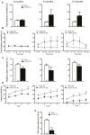
Time-dependent appearance of motor deficits in TDP-43-WT mice. (A–D) Motor behavior was analyzed at 3, 6 and 12 months off Dox. (A–C) In order to assess general exploratory locomotion in a novel environment, open field test were performed. Mice were placed in a clear (40 cm × 40 cm × 40 cm) arena, and a 20 min session was used. (A) Total distance traveled in the open field chamber. An increased trend at 6 months became significant at 12 months of Tg expression (*p < 0.05 significantly different from control group, Student’s t test). (B) Open field time bin (segments of 5 min) analysis of total distance traveled show a significant difference at 12 months after Tg expression (*p < 0.05; **p < 0.01 significantly different from control group, repeated-measures two-way ANOVA/Bonferroni post hoc test). (C) Relative center distance during the open field session show no significant differences at any time post Tg induction. (D) Accelerated rotarod test. TDP-43-WT mice display impaired coordination and balance at 3, 6 and 12 months after Tg induction (*p < 0.05; **p < 0.01; ***p < 0.001 significantly different from control group, repeated-measures two-way ANOVA/Bonferroni post hoc test). (E) Grip strength was evaluated at 12 months after Tg expression using the hanging wire grip test. The latency to fall off the wire lid was quantified using a 60 s cutoff time. TDP-43-WT mice show significant deficits in grip strength (***p < 0.001 significantly different from control group, Student’s t test). Number of animals is indicated in parentheses or inside plot bars. Data represent mean ± SEM.

References

    1. Alami N. H., Smith R. B., Carrasco M. A., Williams L. A., Winborn C. S., Han S. S., et al. . (2014). Axonal transport of TDP-43 mRNA granules is impaired by ALS-causing mutations. Neuron 81, 536–543. 10.1016/j.neuron.2013.12.018 - DOI - PMC - PubMed
    1. Alfieri J. A., Pino N. S., Igaz L. M. (2014). Reversible behavioral phenotypes in a conditional mouse model of TDP-43 proteinopathies. J. Neurosci. 34, 15244–15259. 10.1523/JNEUROSCI.1918-14.2014 - DOI - PMC - PubMed
    1. Amlie-Wolf A., Ryvkin P., Tong R., Dragomir I., Suh E., Xu Y., et al. . (2015). Transcriptomic changes due to cytoplasmic TDP-43 expression reveal dysregulation of histone transcripts and nuclear chromatin. PLoS One 10:e0141836. 10.1371/journal.pone.0141836 - DOI - PMC - PubMed
    1. Ayala Y. M., De Conti L., Avendaño-Vázquez S. E., Dhir A., Romano M., D’Ambrogio A., et al. . (2011). TDP-43 regulates its mRNA levels through a negative feedback loop. EMBO J. 30, 277–288. 10.1038/emboj.2010.310 - DOI - PMC - PubMed
    1. Baralle M., Buratti E., Baralle F. E. (2013). The role of TDP-43 in the pathogenesis of ALS and FTLD. Biochem. Soc. Trans. 41, 1536–1540. 10.1042/BST20130186 - DOI - PubMed

LinkOut - more resources