. 2016 Dec 20:7:496.
doi: 10.3389/fphar.2016.00496. eCollection 2016.

Antinociceptive Effects of AGAP, a Recombinant Neurotoxic Polypeptide: Possible Involvement of the Tetrodotoxin-Resistant Sodium Channels in Small Dorsal Root Ganglia Neurons

Affiliations

Antinociceptive Effects of AGAP, a Recombinant Neurotoxic Polypeptide: Possible Involvement of the Tetrodotoxin-Resistant Sodium Channels in Small Dorsal Root Ganglia Neurons

Chun-Li Li et al. Front Pharmacol. .

Abstract

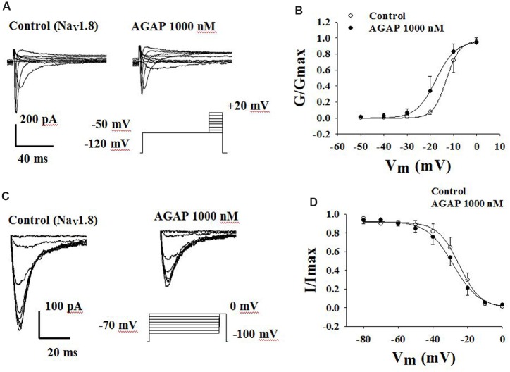
Antitumor-analgesic peptide (AGAP) is a novel recombinant polypeptide. The primary study showed that AGAP 1.0 mg/kg exhibited strong analgesic and antitumor effects. The tail vein administration of AGAP potently reduced pain behaviors in mice induced by intraplantar injection of formalin or intraperitoneal injection of acetic acid, without affecting basal pain perception. To further assess the mechanisms of AGAP, the effects of AGAP on sodium channels were assessed using the whole-cell patch clamp recordings in dorsal root ganglia (DRG) neurons. The results showed that AGAP (3-1000 nM) inhibited the sodium currents in small-diameter DRG neurons in a dose-dependent manner. 1000 nM AGAP could inhibit the current density-voltage relationship curve of sodium channels in a voltage-dependent manner and negatively shift the activation curves. 1000 nM AGAP could reduce the tetrodotoxin-resistant (TTX-R) sodium currents by 42.8% in small-diameter DRG neurons. Further analysis revealed that AGAP potently inhibited NaV1.8 currents by 59.4%, and negatively shifted the activation and inactivation kinetics. 1000 nM AGAP also reduced the NaV1.9 currents by 33.7%, but had no significant effect on activation and inactivation kinetics. Thus, our results demonstrated that submicromolar concentrations of AGAP inhibited TTX-R sodium channel in rat small-diameter DRG neurons. It is concluded that these new results may better explain, at least in part, the analgesic properties of this polypeptide.

Keywords: Nav1.8; Nav1.9; Voltage-gated sodium channels; pain; scorpion toxins.


Figures

FIGURE 1
FIGURE 1
Effect of Antitumor-analgesic peptide (AGAP) on acetic acid-induced writhing test in mice. After intravenously injection of AGAP, mice received an intraperitoneal injection of 0.8% acetic acid, and abdominal writhes were counted for 30 min. AGAP dose-dependently inhibited the total number of writhes. ∗∗p < 0.01, ∗∗∗p < 0.001 compared with control group. It is no significance of comparison between morphine and 1.0 mg/kg AGAP (p > 0.05). Data are shown as the mean ± SEM (n = 12 per group). P values are from one-way ANOVA followed by Tukey’s test.
FIGURE 2
FIGURE 2
Reaction time of animals in hot plate test: 0, 30, 60, 90, and 120 min after treatment of AGAP and morphine. Animals were pretreated with vehicle, morphine (5 mg/kg), AGAP (0.25, 0.5, and 1.0 mg/kg) prior to the tests at 55°C. Each column represents the mean with X ± SEM. for twelve mice in each group. The symbols denote the significance levels: ∗∗p < 0.01, ∗∗∗p < 0.001 compared with control group. Morphine and 1.0 mg/kg AGAP were not statistical difference (p > 0.05). Data are shown as the mean ± SEM (n = 12 per group). P values are from two-way ANOVA followed by Bonferroni test.
FIGURE 3
FIGURE 3
Effect of AGAP against formalin induced licking and flinching in mice. AGAP at concentrations ranging from 0.25 to 1.0 mg/kg was administered intravenously 20 min before formalin injection. Morphine was administered at a dose of 2 mg/kg i.p. 30 min before formalin injection. The total time spent on licking and flinching of the affected paw was measured in the first (0–5 min) phase and the second (15–30 min) phase after intraplantar injection of formalin. AGAP dose-dependently attenuated the formalin-induced licking and flinching responses in both two phases. ###p < 0.001 compared with control group; p < 0.05, ∗∗∗p < 0.001 compared with formalin group. Data are shown as the mean ± SEM (n = 12 per group). P values are from two-way ANOVA followed by Bonferroni test.
FIGURE 4
FIGURE 4
Expression profiles of NaV1.8 and NaV1.9 in DRG neurons (×200). Double immunofluorescent labeling of L4-6 DRG neurons by anti-NaV1.8 (red) and anti-NF200 (green) or anti-NaV1.9 (red) and anti-NF200 (green) antibodies.
FIGURE 5
FIGURE 5
AGAP inhibits voltage-gated sodium current in primary cultured and acutely isolated small-diameter neurons of rat. (A,B) Following the blockade of calcium and potassium channels, depolarizing voltage commands from -80 to +50 mV from a holding potential of -90 mV elicited fast inward currents. The inward current responses were significantly reduced after the application of AGAP (1000 nM) in cultured and acutely isolated small-diameter DRG neurons. (C) The percentage inhibition for INa was 46.9 ± 8.7 and 41.1 ± 6.2% in cultured and acutely isolated small-diameter DRG neurons, respectively (p < 0.05, P values are from unpaired t-test, n = 15 and n = 19, respectively).
FIGURE 6
FIGURE 6
Effect of AGAP on NaV channel currents in small-diameter DRG neurons. (A) Current density-voltage relationship obtained by plotting current density as a function of test potential under control conditions and after application of 1000 nM AGAP. (n = 7). (B,C) The graph shows the dose–response relationship of the effect of AGAP in acutely isolated small-diameter DRG neurons (P < 0.05, ∗∗P < 0.01, ∗∗∗P < 0.001, P values are from unpaired t-test. n = 8–13). (D) Effect of AGAP on steady-state activation kinetics of NaV channels. Data points were fitted with the Boltzmann equation. V1/2 was -19.1 ± 0.3 mV under control conditions and -25.0 ± 0.7 mV in the presence 1000 nM of AGAP (P < 0.05, P values are from unpaired t-test. n = 7); slope factors were 3.2 ± 0.3 and 2.7 ± 0.3 mV, respectively.
FIGURE 7
FIGURE 7
Effect of AGAP on the TTX-R sodium currents in small-diameter DRG neurons. (A) Representative traces showing the depression of 1000 nM TTX on NaV channel currents in rat small diameter DRG neuron. (B) Current density-voltage relationship obtained by plotting current density as a function of test potential under control conditions and after application of 1000 nM TTX (n = 6). (C) Representative traces showing the depression of AGAP on TTX-R currents in a neuron. (D) 1000 nM AGAP inhibited TTX-R currents by 42.8% (P < 0.05, P values are from unpaired t-test, n = 9). (E) Representative traces showing the depression of AGAP on NaV1.8 currents in a neuron. (F) At a concentration of 1000 nM, AGAP potently inhibited NaV1.8, decreasing current amplitude by 59.4% (P < 0.01, P values are from unpaired t-test. n = 5). (G) Representative traces showing the depression of AGAP on NaV1.9 currents in a neuron. (H) 1000 nM AGAP reduced NaV1.9 currents by 33.7% (P < 0.05, P values are from unpaired t-test. n = 9).
FIGURE 8
FIGURE 8
Effect of AGAP on the dynamic function of NaV1.8 channels. (A,B) AGAP shifts the conductance-voltage relationship to more negative potentials. (A) Representative steady-state activation of NaV1.8 under the control conditions and in the presence of 1000 nM AGAP. (B) Normalized activation kinetics determined before and after the application of AGAP. V1/2 was -13.0 ± 0.2 mV in control conditions and -17.6 ± 0.4 mV in the presence of 1000 nM AGAP (P < 0.05, n = 5). Slope factors were 2.8 ± 0.1 and 4.2 ± 0.3 mV, respectively (P < 0.05, n = 5). P values are from unpaired t-test. (C,D) AGAP had no effect on the voltage-dependence of steady-state inactivation. (C) Representative steady-state inactivation of NaV1.8 under the control conditions and in the presence of 1000 nM AGAP. (D) Normalized peak currents were plotted against membrane potential and Boltzmann equation was used to fit data. V1/2 was -25.0 ± 0.3 mV in control conditions and -25.4 ± 0.6 mV in the presence of 1000 nM AGAP. Slope factors were -5.5 ± 0.3 and -6.0 ± 0.5 mV, respectively (P > 0.05, P values are from unpaired t-test. n = 5).
FIGURE 9
FIGURE 9
Effect of AGAP on the dynamic function of NaV1.9 channels. 1000 nM AGAP did not significantly affect the voltage-dependence of channel activation or steady-state inactivation. (A,B) Effect of AGAP on steady-state activation kinetics of NaV1.9 currents. V1/2 was -38.4 ± 1.5 mV in control conditions and -38.2 ± 3.7 mV in the presence of 1000 nM AGAP. Slope factors were 6.1 ± 0.5 and 7.8 ± 1.1 mV, respectively (n = 7). (C,D) Steady-state inactivated of Nav1.9 in the absence and presence of AGAP. V1/2 for inactivation was -67.9 ± 2.2 mV in control conditions and -69.6 ± 2.5 mV in the presence of 1000 nM AGAP. Slope factors were -11.0 ± 1.8 and -12.5 ± 2.0 mV, respectively (P > 0.05, P values are from unpaired t-test. n = 6).

References

    1. Akopian A. N., Sivilotti L., Wood J. N. (1996). A tetrodotoxin-resistant voltage-gated sodium channel expressed by sensory neurons. Nature 379 257–262. 10.1038/379257a0 - DOI - PubMed
    1. Amaya F., Decosterd I., Samad T. A., Plumpton C., Tate S., Mannion R. J., et al. (2000). Diversity of expression of the sensory neuron-specific TTX-resistant voltage-gated sodium ion channels SNS and SNS2. Mol. Cell. Neurosci. 15 331–342. 10.1006/mcne.1999.0828 - DOI - PubMed
    1. Amaya F., Wang H., Costigan M., Allchorne A. J., Hatcher J. P., Egerton J., et al. (2006). The voltage-gated sodium channel Na(v)1.9 is an effector of peripheral inflammatory pain hypersensitivity. J. Neurosci. 26 12852–12860. 10.1523/JNEUROSCI.4015-06.2006 - DOI - PMC - PubMed
    1. Amir R., Argoff C. E., Bennett G. J., Cummins T. R., Durieux M. E., Gerner P., et al. (2006). The role of sodium channels in chronic inflammatory and neuropathic pain. J. Pain 7(5 Suppl. 3), S1–S29. 10.1016/j.jpain.2006.01.444 - DOI - PubMed
    1. Araújo I. W., Rodrigues J. A., Quindere A. L., Silva J., Maciel G., Rebeiro K. A., et al. (2016). Analgesic and anti-inflammatory actions on bradykinin route of a polysulfated fraction from alga Ulva lactuca. Int. J. Biol. Macromol. 92 820–830. 10.1016/j.ijbiomac.2016.07.094 - DOI - PubMed

LinkOut - more resources