Review
. 2016 Dec 21:7:594.
doi: 10.3389/fimmu.2016.00594. eCollection 2016.

Targeting Innate-Like T Cells in Tuberculosis

Affiliations
Review

Targeting Innate-Like T Cells in Tuberculosis

Shouxiong Huang. Front Immunol. .

Abstract

Peptide-specific conventional T cells have been major targets for designing most antimycobacterial vaccines. Immune responses mediated by conventional T cells exhibit a delayed onset upon primary infection and are highly variable in different human populations. In contrast, innate-like T cells quickly respond to pathogens and display effector functions without undergoing extensive clonal expansion. Specifically, the activation of innate-like T cells depends on the promiscuous interaction of highly conserved antigen-presenting molecules, non-peptidic antigens, and likely semi-invariant T cell receptors. In antimicrobial immune responses, mucosal-associated invariant T cells are activated by riboflavin precursor metabolites presented by major histocompatibility complex-related protein I, while lipid-specific T cells including natural killer T cells are activated by lipid metabolites presented by CD1 proteins. Multiple innate-like T cell subsets have been shown to be protective or responsive in mycobacterial infections. Through rapid cytokine secretion, innate-like T cells function in early defense and memory response, offering novel advantages over conventional T cells in the design of anti-tuberculosis strategies.

Keywords: CD1; MR1; Mycobacterium tuberculosis; antigen presentation; innate-like T cells; lipid; riboflavin metabolites; vaccine.


Figures

Figure 1
Figure 1
Responding kinetics of mucosal-associated invariant T (MAIT) and natural killer T (NKT) cells. (A) In vivo responding kinetics were hypothesized based on the presence of mycobacterial antigen-specific CD8+ T cells in lung tissues (3), the ability of MAIT cells to inhibit Bacillus Calmette–Guérin (BCG) growth in lung tissues (80), and the ability of transferred iNKT to inhibit M. tuberculosis growth in lung tissues (81). (B) In vitro-responding kinetics were estimated according to the acquisition of cytolytic function by CD8+ T cells upon in vitro peptide stimulation (77), cytokine production by polyclonal MAIT cells upon stimulation with BCG-infected macrophages (80), and cytokine production by tetramer-isolated human polyclonal NKT cells upon antigen-specific activation (82).
Figure 2
Figure 2
Ligand-binding and TCR contact of MR1 protein inferred from mutagenesis (84) and structural (34) studies. (A) Mutated mouse MR1 residues (84) are annotated on a human MR1 crystal structure (hMR1-RL-6-Me-7-OH) (34) to show the resulted functional impact on MR1 surface expression and mucosal-associated invariant T (MAIT) cell activation through predicted ligand interactions (green). Red: mutated residues on the β-sheet or proximal regions showing low MR1 expression and/or inhibited MAIT cell activation; gray: ligand-interacting residues not mutated. (B) Structural interactions between MR1 protein and the ligand RL-6-Me-7-OH (green) (34). (C) Mutated mouse MR1 residues (84) are annotated on hMR1 crystal structure (34) to show the resulted functional impact on T cell activation through predicted interaction with MAIT cell TCR. Red: mutated residues on helical regions showing inhibited MAIT cell activation; gray: mutated helical residues not showing functional impact. (D) Surface MR1 residues that structurally interact with corresponding complementarity-determining regions (CDRs) are shown with the same color (34).
Figure 3
Figure 3
MR1 and CD1 ligand-binding clefts. The crystal structures of MR1 (A) (34), CD1a (B) (58), and CD1d (C) (134) molecules are shown with associated ligands (green) in comparison to the HLA-B2705 protein (D) (135). The positions of the portals for the MR1 and CD1 ligand-binding clefts are annotated. The shapes and relative sizes of the MR1 and CD1 ligand-binding clefts are also shown with ribbon view.
Figure 4
Figure 4
Docking mode of T cell receptors to MR1, CD1, and HLA-B2705 proteins. Tri-molecular interaction among an antigen-presenting molecule, an antigen, and a TCR in the activation of unconventional T cells is represented with the tri-molecular complexes of (A) human MR1-ribityllumazine–mucosal-associated invariant T cell TCR (34), (B) human CD1a–lysophosphatidylcholine–TCR of BK6 T cell (58), (C) mouse CD1d–α-galactosyl diacylglycerol (α-GalDAG)–iNKT TCR (134), and (D) HLA-B2705-KK10 peptide from human immunodeficiency virus Gag protein-TCR (135). The color-coated residues annotate the regions on the surface of antigen-presenting molecules to interact with CDR regions of TCRs (bluish colors for TCRα interactions and reddish colors for TCRβ interactions). TCRs from unconventional T cells more dominantly contact the surfaces of MR1 and CD1 proteins than the metabolite antigens. However, TCRs from conventional T cells recognize both HLA proteins and peptide antigens.
Figure 5
Figure 5
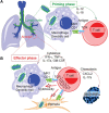
Proposed priming and effector phases of innate-like T cell responses in tuberculosis. (A) The priming phase may occur in the lymph nodes of lung tissues. Naïve or precursor innate-like T cells are activated through interaction with MR1 and CD1 proteins and/or stimulation of cytokines. (B) Activated innate-like T cells migrate to infected tissues, such as alveolar regions, to perform cytotoxic function and secrete cytokines and chemokines in anti-M. tuberculosis effector response.

Similar articles

Cited by

References

    1. Flynn JL, Chan J. Immunology of tuberculosis. Annu Rev Immunol (2001) 19:93–129.10.1146/annurev.immunol.19.1.93 - DOI - PubMed
    1. Cooper AM. Cell-mediated immune responses in tuberculosis. Annu Rev Immunol (2009) 27:393–422.10.1146/annurev.immunol.021908.132703 - DOI - PMC - PubMed
    1. Behar SM. Antigen-specific CD8(+) T cells and protective immunity to tuberculosis. Adv Exp Med Biol (2013) 783:141–63.10.1007/978-1-4614-6111-1_8 - DOI - PMC - PubMed
    1. Orme IM, Collins FM. Adoptive protection of the Mycobacterium tuberculosis-infected lung. Dissociation between cells that passively transfer protective immunity and those that transfer delayed-type hypersensitivity to tuberculin. Cell Immunol (1984) 84(1):113–20.10.1016/0008-8749(84)90082-0 - DOI - PubMed
    1. Tascon RE, Stavropoulos E, Lukacs KV, Colston MJ. Protection against Mycobacterium tuberculosis infection by CD8+ T cells requires the production of gamma interferon. Infect Immun (1998) 66(2):830–4. - PMC - PubMed

LinkOut - more resources