Review
. 2016 Nov;8(11):3401-3413.
doi: 10.21037/jtd.2016.11.24.

Tracheobronchial tumors

Affiliations
Review

Tracheobronchial tumors

Ruza Stevic et al. J Thorac Dis. 2016 Nov.

Abstract

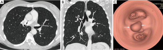
Tumors of trachea and bronchi are uncommon and can occur in the form of benign or low- and high-grade malignant tumors. Although tracheobronchial tumors (TBTs) represent only 0.6% of all pulmonary tumors, they are clinically significant. Delays in diagnosis of these tumors commonly occur because the signs and symptoms caused by these tumors are nonspecific and chest radiographs are often considered unremarkable. Therefore, novel radiological techniques and better access to flexible bronchoscopy enable detection of larger number of TBT. The purpose of this article is to provide a review of tracheal and bronchial tumors and discuss significant aspects of the different TBT with focus on clinical manifestations and diagnostic procedures.

Keywords: Trachea; bronchi; diagnosis; tumor.


Conflict of interest statement

The authors have no conflicts of interest to declare.

Figures

Figure 1
Figure 1
Flow-volume curve in the patient with tracheal tumor.
Figure 2
Figure 2
Axial (A) and coronal MPR (B) images shows tumor mass in left main bronchus (arrows). Virtual bronchoscopy (C) of the same patient shows a tan spherical, smooth-surfaced tumor in left main bronchus (arrows).
Figure 3
Figure 3
Squamous cell carcinoma of trachea. Axial (A) and coronal MPR (B) images show eccentric soft tissue mass with regular margins involving trachea with mediastinal extension (arrow in A,B). PET CT shows high FDG uptake (arrow) suggesting malignancy (C).
Figure 4
Figure 4
Squamous cell lung carcinoma. Contrast enhanced axial (A) and coronal MPR (B) images show obstructing tumor mas in right lower lobe (arrow in A,B) with distal atelectasis(C).
Figure 5
Figure 5
Chest CT of patient with carcinoid tumor. CT scan shows an oval tumor inside the left upper lobe bronchus arrows) and hyperlucency of the same lobe.
Figure 6
Figure 6
Typical carcinoid tumor. Contrast-enhanced axial (A), MPR (B) images and coronal reconstructed MinIP (C) show a well-defined, soft tissue nodule (arrow) in the left main bronchus.
Figure 7
Figure 7
Tracheal metastasis from colorectal cancer. Axial (A) and coronal MPR (B) images show soft tissue mass causing reduction of tracheal lumen (arrows in A,B). PET CT (C) image shows increased FDG uptake in lesion (arrow).
Figure 8
Figure 8
Endobronchial hamartoma presented as endobronchial mass in lingular bronchus (arrow) and distal pneumonia on axial CT scan.
Figure 9
Figure 9
Endobronchial lipoma. Axial CT scans in soft tissue (A) and lung window setting (B) shows round tumor in posterior segmental bronchus of the right lower lobe and signs of inflammation.
Figure 10
Figure 10
Bronchial papilloma. Axial (A), coronal MPR image (B) show a smooth polypoidal soft tissue mass arising from the anterior wall of right lower bronchus (arrow). Virtual bronchoscopy (C) shows the intraluminal mass lesion with smooth surface (arrow).

References

    1. Xu LT, Sun ZF, Li ZJ, et al. Clinical and pathologic characteristics in patients with tracheobronchial tumor: Report of 50 patients. Ann Thorac Surg 1987;43:276-8. 10.1016/S0003-4975(10)60611-X - DOI - PubMed
    1. Stevic R, Milenkovic B, Stojsic J, et al. Clinico-radiological characteristics of tracheobronchial tumors: Report of 65 cases. Ann Acad Med Singapore 2012;41:205-11. - PubMed
    1. Wilson RW, Kirejczyk W. Pathological and radiological correlation of endobronchial neoplasms: Part I, benign tumors. Ann Diagn Pathol 1997;1:31-46. 10.1016/S1092-9134(97)80007-X - DOI - PubMed
    1. Wilson RW, Frazier AA. Pathological-radiological correlations: pathological and radiological correlation of endobronchial neoplasms: part II, malignant tumors. Ann Diagn Pathol 1998;2:31-4. 10.1016/S1092-9134(98)80033-6 - DOI - PubMed
    1. Ko JM, Jung JI, Park SH, et al. Benign tumors of the tracheobronchial tree: CT-pathologic correlation. AJR Am J Roentgenol 2006;186:1304-13. 10.2214/AJR.04.1893 - DOI - PubMed

LinkOut - more resources