

Initial and sustained brain responses to threat anticipation in blood-injection-injury phobia
- PMID: 28066706
- PMCID: PMC5200881
- DOI: 10.1016/j.nicl.2016.12.015
Initial and sustained brain responses to threat anticipation in blood-injection-injury phobia
Abstract
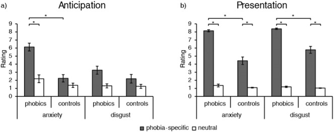
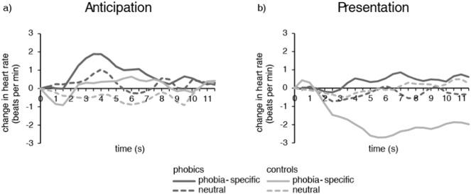
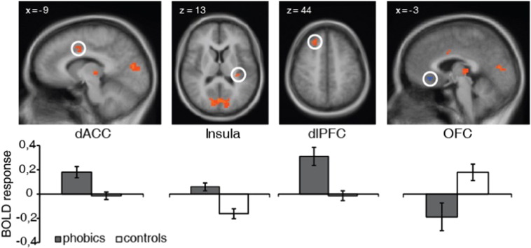
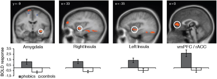
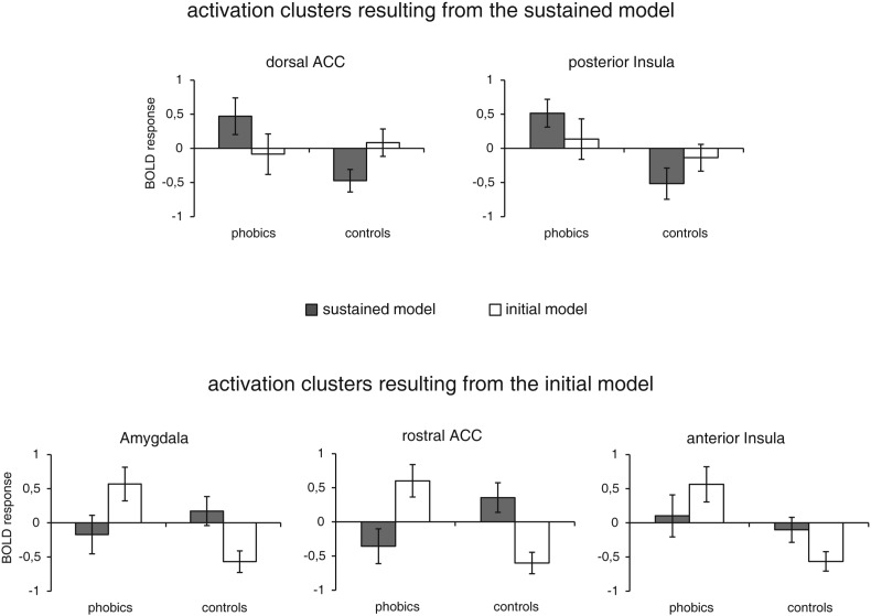
Blood-injection-injury (BII) phobia differs from other subtypes of specific phobia in that it is associated with elevated disgust-sensitivity as well as specific autonomic and brain responses during processing of phobia-relevant stimuli. To what extent these features play a role already during threat anticipation is unclear. In the current fMRI experiment, 16 female BII phobics and 16 female healthy controls anticipated the presentation of phobia-specific and neutral pictures. On the behavioral level, anxiety dominated the anticipatory period in BII phobics relative to controls, while both anxiety and disgust were elevated during picture presentation. By applying two different models for the analysis of brain responses to anticipation of phobia-specific versus neutral stimuli, we found initial and sustained increases of activation in anterior cingulate cortex (ACC), insula, lateral and medial prefrontal cortex (PFC), thalamus and visual areas, as well as initial activation in the amygdala for BII phobics as compared to healthy controls. These results suggest that BII phobia is characterized by activation of a typical neural defense network during threat anticipation, with anxiety as the predominant emotion.
Keywords: Amygdala; Anterior cingulate cortex; Bed nucleus of the stria terminalis (BNST); Insula; fMRI.
Figures
References
-
- American Psychiatric Association . 2000. Diagnostic and Statistical Manual of Mental Disorders, 4th Edition (DSM-IV) (Washington, D.C.)
-
- Amodio D.M., Frith C.D. Meeting of minds: the medial frontal cortex and social cognition. Nat. Rev. Neurosci. 2006;7:268–277. - PubMed
-
- Andrews G., Freed S., Teesson M. Proximity and anticipation of a negative outcome in phobias. Behav. Res. Ther. 1994;32:643–645. - PubMed
-
- Aue T., Hoeppli M.-E. Evidence for an encounter expectancy bias in fear of spiders. Cognit. Emot. 2011;26:727–736. - PubMed
Publication types
MeSH terms
Supplementary concepts
LinkOut - more resources
Full Text Sources
Other Literature Sources
Medical
Miscellaneous
