. 2016 Aug 26;1(8):975-978.
doi: 10.1021/acssensors.6b00396. Epub 2016 Aug 10.

pH-Dependent Cellular Internalization of Paramagnetic Nanoparticle

Affiliations

pH-Dependent Cellular Internalization of Paramagnetic Nanoparticle

Branislava Janic et al. ACS Sens. .

Abstract

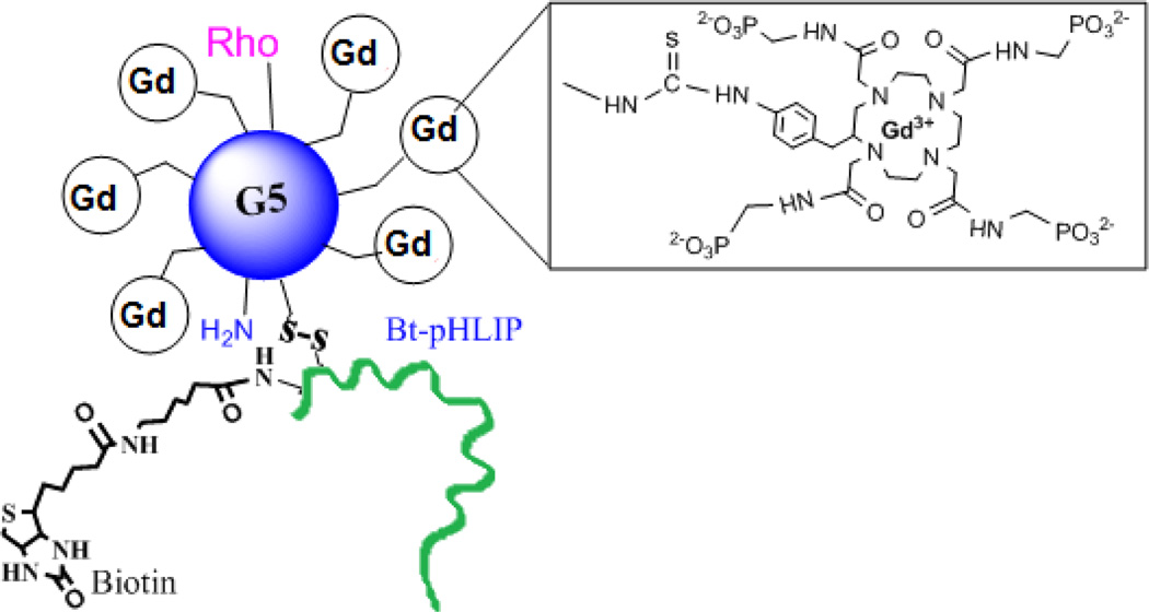
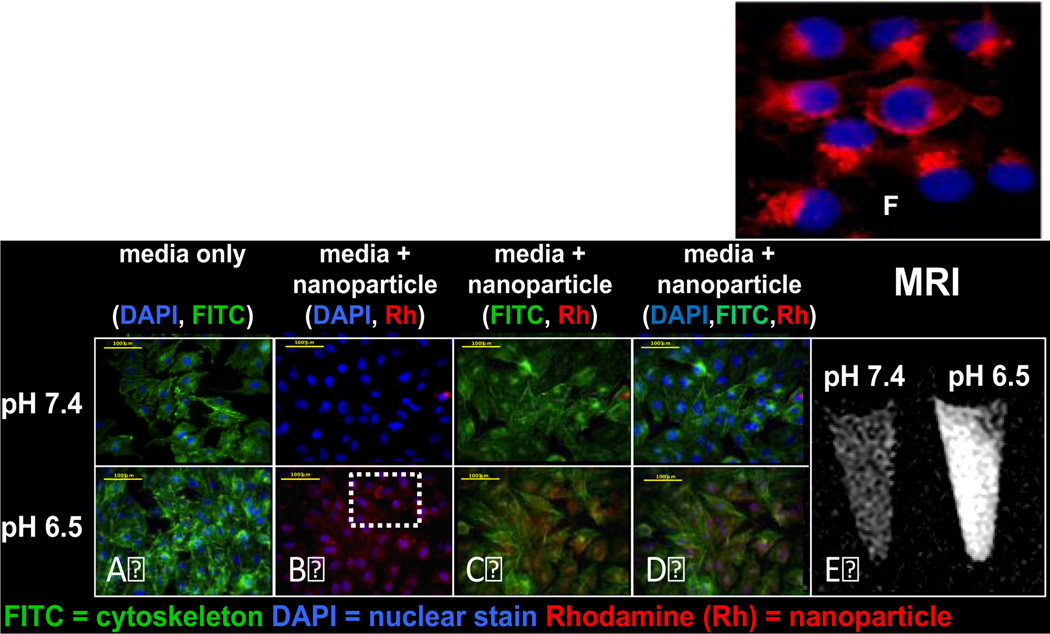
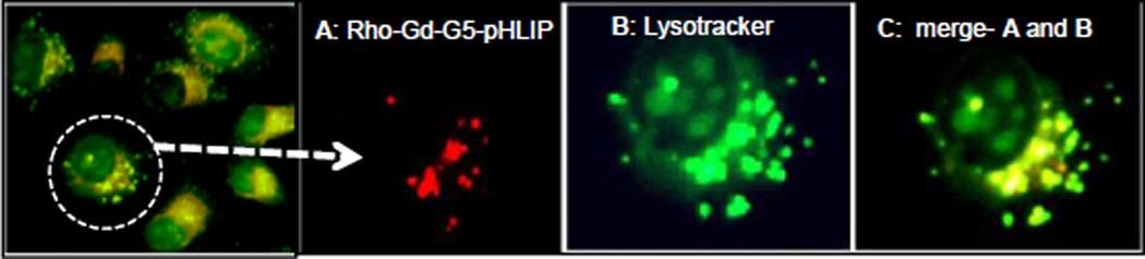
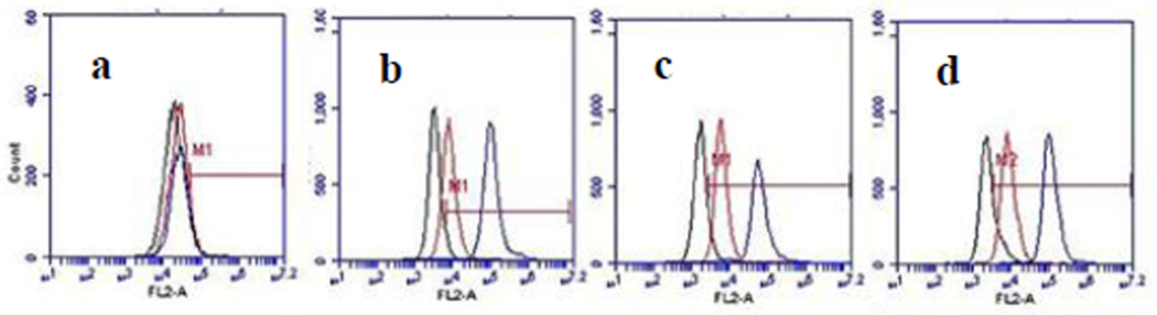
A hallmark of the tumor microenvironment in malignant tumor is extracellular acidosis, which can be exploited for targeted delivery of drugs and imaging agents. A pH sensitive paramagnetic nanoaparticle (NP) is developed by incorporating GdDOTA-4AmP MRI contrast agent and pHLIP (pH Low Insertion Peptide) into the surface of a G5-PAMAM dendrimer. pHLIP showed pH-selective insertion and folding into cell membranes, but only in acidic conditions. We demonstrated that pHLIP-conjugated Gd44-G5 paramagnetic nanoparticle binds and fuses with cellular membrane at low pH, but not at normal physiological pH, and that it promotes cellular uptake. Intracellular trafficking of NPs showed endosomal/lysosomal path ways.

Keywords: Cell internalization; MRI; intracellular trafficking; pH sensing probe; paramagnetic nanoparticle.


Figures

Figure 1
Figure 1
Schematic view of Gd3+ chelated with 1,4,7,10-tetraaza-cyclododecane-1,4,7,10-tetraaminophosphonate (DOTA-4AmP8-) in a Gd44-G5 PAMAM dendrimer with rhodamine (Rho) and Bt-pHLIP conjugation. Gd44-G5 is linked with Bt-pHLIP through sulphur-sulphur bond.
Figure 2
Figure 2
In vitro fluorescence microscopy of MDA-MB-231 cells incubated for 3 hours in media containing rhodamine B conjugated Gd44-G5-pHLIP3 dendrimer at pH 7.4 and at pH 6.5 at a concentration of 7.1 µM with respect to rhodamine B. Nuclei were visualized with DAPI (blue fluorescence). Cyto-skeleton was visualized by FITC CytoPainter F-Actin specific dye (green fluorescence). Intracellular uptake was visualized by red fluorescence of Rhodamine conjugated to the nanoparticle (see B). Scale bars = 100 µm. Panel A showing cells incubated in media only. Images of cells incubated in the presence of media and nanoparticle were captured using DAPI and Rh filters (B), FITC and Rh filters (C) and DAPI, FITC and Rh filters (D). Cytoskeleton was visualized by FITC Cyto-Painter F-Actin specific dye. Panel 2E: In-vitro NP specificity quantified by MRI. T1- maps of gel phantoms containing MDA-MB-231 cells incubated with NP at pH 6.5 and 7.4. Panel 2F: Enlarged image of perinuclear distribution of Rho-Gd44-G5-ss-pHLIP3 from overlaid images of dapi and nanoparticle (B).
Figure 3
Figure 3
In vitro fluorescence microscopy images of MDA-MB-231 live cell incubated for 2 hours in media containing rhodamine B conjugated Gd-G5—ss-Bt-pHLIP3 dendrimer at pH 6.5. Nano-particle is red (A) and lysotracker green, detecting endosomes and lysosomes is shown in green (B). Overlay between (A) and (B) is represented in yellow (C). Overlaid images are shown in the far left and far right panel.
Figure 4
Figure 4
Flow cytometry of the cell entry dynamics of Rho-Gd44-G5-ss-Bt-pHLIP3 conjugate (particle is attached to C-terminus of the peptide) in MSC (a) (mesenchymal stem cell), U251 (b), MDA-MB-231 (c) and U87 (d) cell lines. The log of rhodamine adsorption intensity (FL1-H on X axis is plotted against the number of cells (counts on y axis). No cellular uptake of the nanoparticle in normal MSC (a) was observed for both pH (6.5 and 7.4). In contrast, the cellular uptake of the nanaoparticle in all three cancer cell lines (b, c & d) is evident at lower pH 6.5. (black line) Control (no probe at pH 7.4); (red line) nanoparticle at pH 7.4; (blue line) nanoparticle at pH 6.5.

Similar articles

Cited by

References

    1. Srinivas PR, Kramer BS, Srivastava S. Trends in biomarker research for cancer detection. The lancet oncology. 2001;2(11):698–704. - PubMed
    1. Hanke JH, Webster KR, Ronco LV. Protein biomarkers and drug design for cancer treatments. Eur J Cancer Prev. 2004;13(4):297–305. - PubMed
    1. Bild AH, Yao G, Chang JT, et al. Oncogenic pathway signatures in human cancers as a guide to targeted therapies. Nature. 2006;439(7074):353–357. - PubMed
    1. Poon Z, Chang D, Zhao X, Hammond PT. Layer-by-layer nanoparticles with a pH-sheddable layer for in vivo targeting of tumor hypoxia. ACS nano. 2011;5(6):4284–4292. - PMC - PubMed
    1. Shenoy D, Little S, Langer R, Amiji M. Poly(ethylene oxide)-modified poly(beta-amino ester) nanoparticles as a pH-sensitive system for tumor-targeted delivery of hydrophobic drugs: part 2. In vivo distribution and tumor localization studies. Pharmaceutical research. 2005;22(12):2107–2114. - PMC - PubMed

LinkOut - more resources