. 2017 Jan 9:7:40075.
doi: 10.1038/srep40075.

Physiological Remediation of Cobalt Ferrite Nanoparticles by Ferritin

Affiliations

Physiological Remediation of Cobalt Ferrite Nanoparticles by Ferritin

Jeanne Volatron et al. Sci Rep. .

Abstract

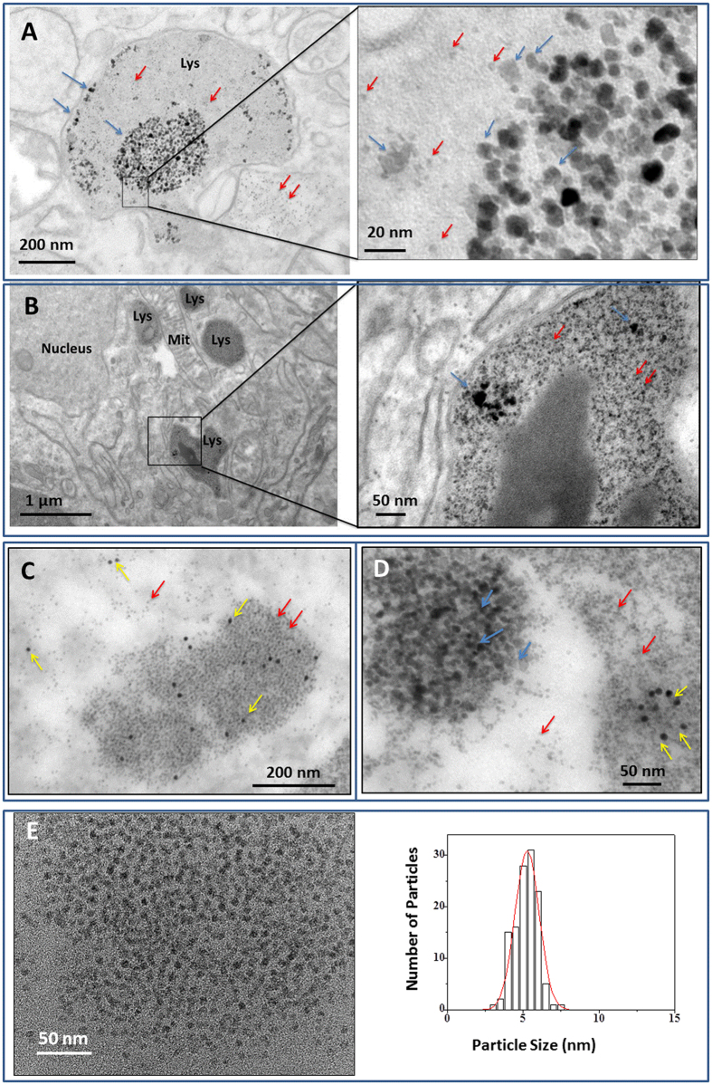
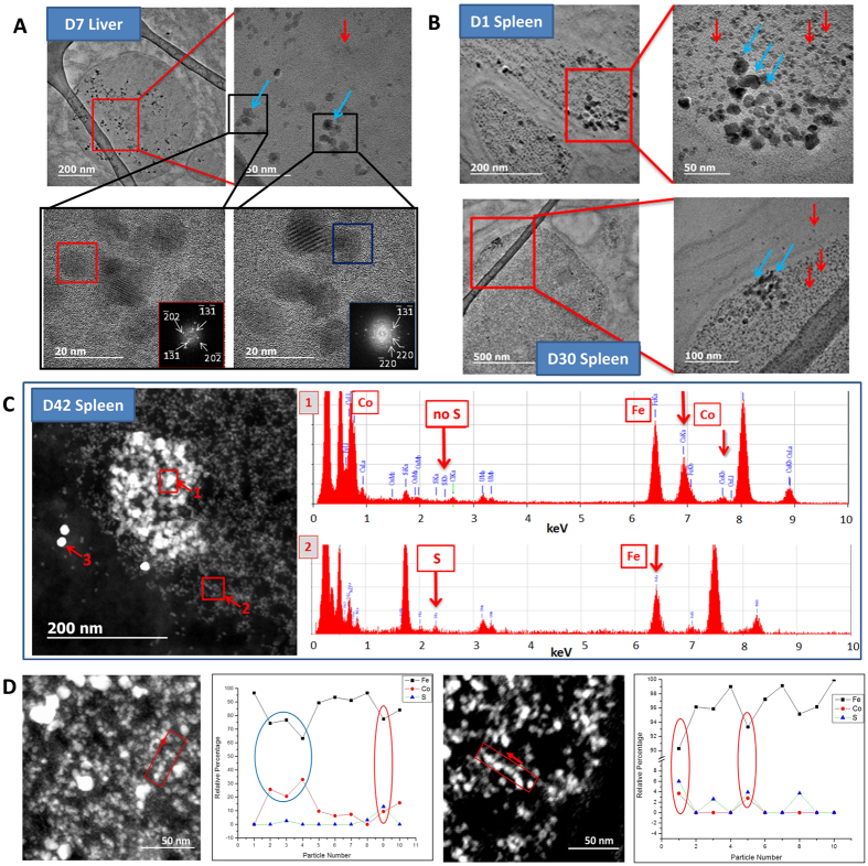
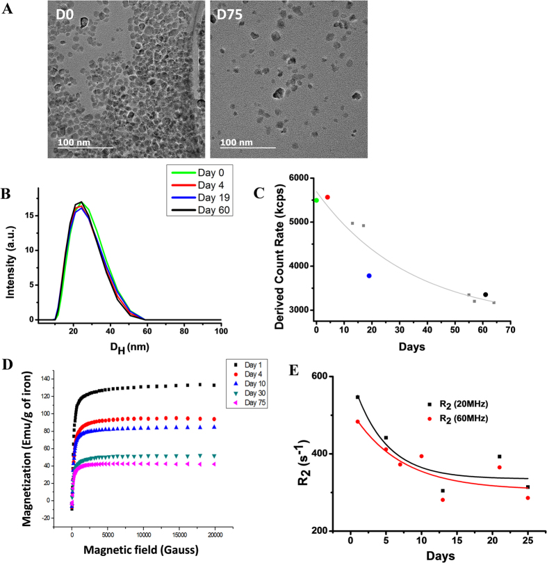
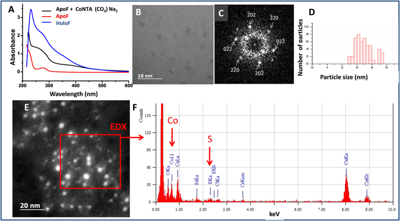
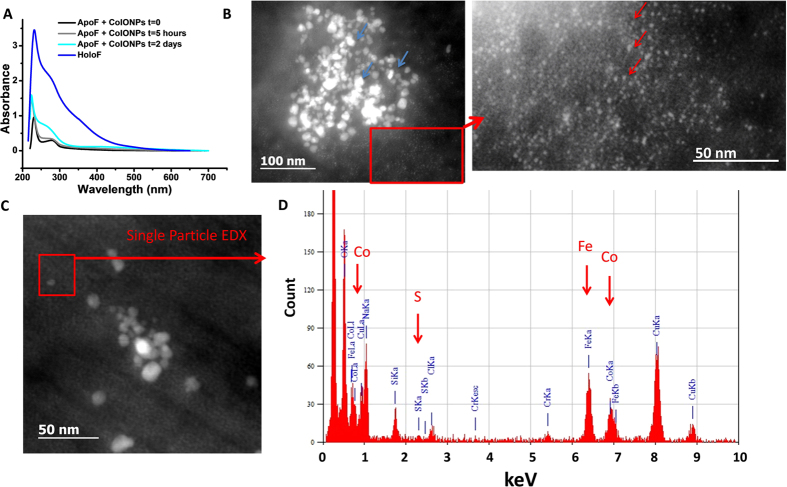
Metallic nanoparticles have been increasingly suggested as prospective therapeutic nanoplatforms, yet their long-term fate and cellular processing in the body is poorly understood. Here we examined the role of an endogenous iron storage protein - namely the ferritin - in the remediation of biodegradable cobalt ferrite magnetic nanoparticles. Structural and elemental analysis of ferritins close to exogenous nanoparticles within spleens and livers of mice injected in vivo with cobalt ferrite nanoparticles, suggests the intracellular transfer of degradation-derived cobalt and iron, entrapped within endogenous protein cages. In addition, the capacity of ferritin cages to accommodate and store the degradation products of cobalt ferrite nanoparticles was investigated in vitro in the acidic environment mimicking the physiological conditions that are present within the lysosomes. The magnetic, colloidal and structural follow-up of nanoparticles and proteins in the lysosome-like medium confirmed the efficient remediation of nanoparticle-released cobalt and iron ions by ferritins in solution. Metal transfer into ferritins could represent a quintessential process in which biomolecules and homeostasis regulate the local degradation of nanoparticles and recycle their by-products.


Figures

Figure 1
Figure 1
TEM micrographs of spleen samples at day 1 (A), day 7 (B) and day 42 (C,D) after intravenous injection of CoIONPs in mice. CoIONPs (indicated with blue arrows) are found in lysosomes (Lys) of macrophages in proximity to iron-filled ferritin proteins (HoloF) (red arrows). In C&D, ferritin proteins are labeled with 10 nm colloidal gold anti-ferritin antibody (yellow arrows). Note the organization of HoloF as large clusters. (E) HoloF aggregates in spleen and corresponding size distribution.
Figure 2
Figure 2. TEM tracking and EDX nano analyses of CoIONPs and ferritins in liver and spleen at different time-points after intravenous injection of CoIONPs.
(A) CoIONPs are identified in the lysosomes of liver macrophages at day 7, by their characteristic size distribution (8.7 ± 2.9 nm) and inverse spinel structure of cobalt ferrite, determined from the FFT of the HRTEM pictures (red and blue squares). (B) CoIONPs (blue arrows) are seen in the neighborhood of numerous HoloF proteins (red arrows) in spleen lysosomes at day 1 (top images) and at day 30 (bottom images). (C) STEM-HAADF micrograph of intralysosomal CoIONPs in spleen at day 42 post injection. The 10 nm monodisperse bright particles (labelled 3) are antiferritin immunogold particles. The less bright and polydisperse particles (square “1”) are the native CoIONPs with a Fe:Co ratio of 2 to 3 and absence of sulfur, as determined by EDX analysis. The small quasi-monodisperse particles correspond to HoloF, characterized by the colocalization of iron and sulfur (Fe:S ratio of about 20). (D) Single particle EDX analyses on spleen samples at day 42 post-injection. The graphs on the right of the STEM-HAADF picture show the relative percentage of Fe, Co and S determined from the point by point EDX analysis of the red quadrant scanned in the arrow direction. Blue oval depicts a native CoIONP particle, while red ovals indicate colocalization of cobalt, iron and sulfur, indicating cobalt uptake by ferritins.
Figure 3
Figure 3. Evolution of citrate-coated CoIONPs in acidic citrate medium.
(A) Representative TEM micrographs at two time-points. (B) Time evolution of the distribution of hydrodynamic sizes and (C) derived count rate in DLS. (D) Time evolution of the field-dependent magnetization curve at 300 K and (E) Time evolution of the transverse NMR relaxation rate R2 at proton Larmor frequencies of 20 MHz and 60 MHz.
Figure 4
Figure 4. Signature of apoferritin filling cobalt from cobalt salt CoNTA (CO3) Na2.
(A) UV-Vis spectrum of ApoF incubated with CoNTA (CO3) Na2 for 22 hours in acidic medium in comparison to ApoF alone and HoloF. Note the growth of the absorbance shoulder at 280 nm indicating the filling of the protein cage. (B) HRTEM of ApoF incubated with CoNTA showing crystalline structures with diffraction pattern (C) characteristic of the cobalt oxide (CoO) along [111] zone axis. (D) Distribution of crystal sizes (mean diameter 3.5 ± 0.6 nm) suggesting a partial filling of the protein cage, in line with UV-Vis spectrum. (E) STEM-HAADF images of ferritin nanocrystals and (F) EDX analysis of the red area, indicated characteristic lines of cobalt and sulfur.
Figure 5
Figure 5. Metal transfer from NPs to apoferritin proteins.
(A) UV-vis spectra of ApoF incubated with CoIONPs in acidic medium for 5 hours and 2 days, in comparison to ApoF and HoloF. The growth of absorbance shoulder at 280 nm indicates metal filling of the protein. (B) STEM-HAADF images of CoIONPs (blue arrows) incubated with ApoF for 2 months. Metal-filled proteins appear as small particles (red arrows). (C,D) Single particle EDX analysis of the red-contoured area in C confirms occupancy of iron, cobalt and sulfur in single ApoF with relative atomic percentages of 64.7%, 28.2% and 7.13% respectively.

References

    1. Feliu N. et al.. In vivo degeneration and the fate of inorganic nanoparticles. Chem. Soc. Rev. 45, 2440–2457 (2016). - PubMed
    1. Caracciolo G., Farokhzad O. C. & Mahmoudi M. Biological Identity of Nanoparticles In Vivo: Clinical Implications of the Protein Corona. Trends Biotechnol. sept 20 (2016) - PubMed
    1. Levy M. et al.. Long term in vivo biotransformation of iron oxide nanoparticles. Biomaterials 32, 3988–3999 (2011). - PubMed
    1. Kolosnjaj-Tabi J. et al.. The One Year Fate of Iron Oxide Coated Gold Nanoparticles in Mice. ACS Nano 9, 7925–7939 (2015). - PubMed
    1. Lartigue L. et al.. Biodegradation of Iron Oxide Nanocubes: High-Resolution In Situ Monitoring. ACS Nano 7, 3939–3952 (2013). - PubMed

Publication types