Review
. 2017 Jan 26;9(4):1356-1392.
doi: 10.1039/c6nr07315h.

Nanocaged platforms: modification, drug delivery and nanotoxicity. Opening synthetic cages to release the tiger

Affiliations
Review

Nanocaged platforms: modification, drug delivery and nanotoxicity. Opening synthetic cages to release the tiger

Mahdi Karimi et al. Nanoscale. .

Abstract

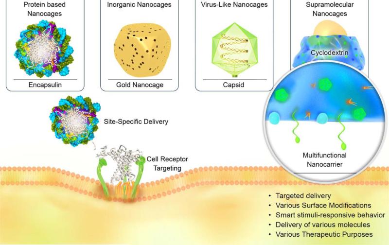
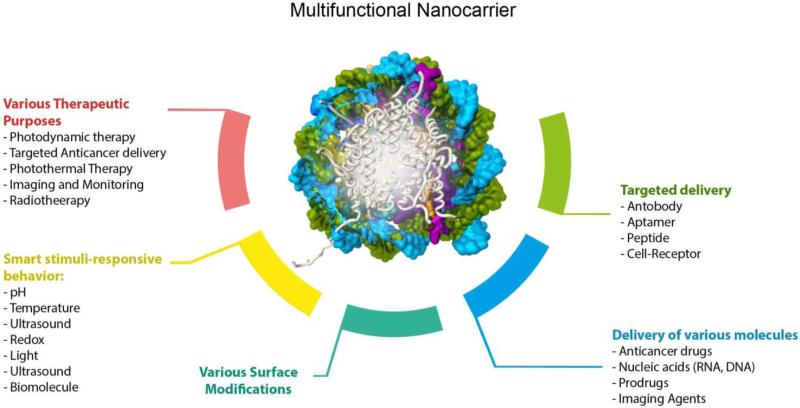
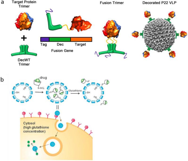
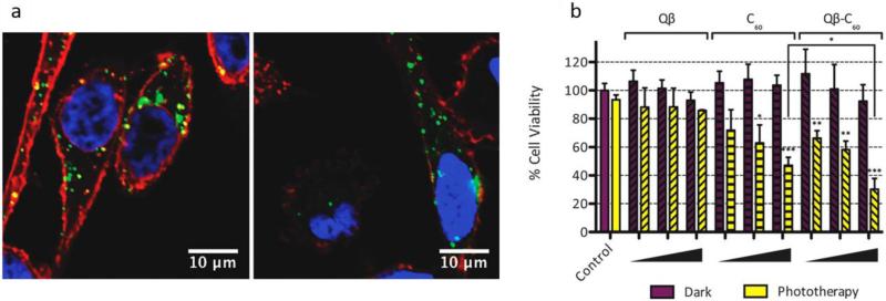
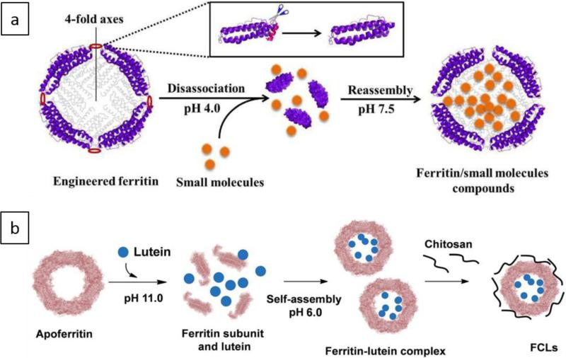
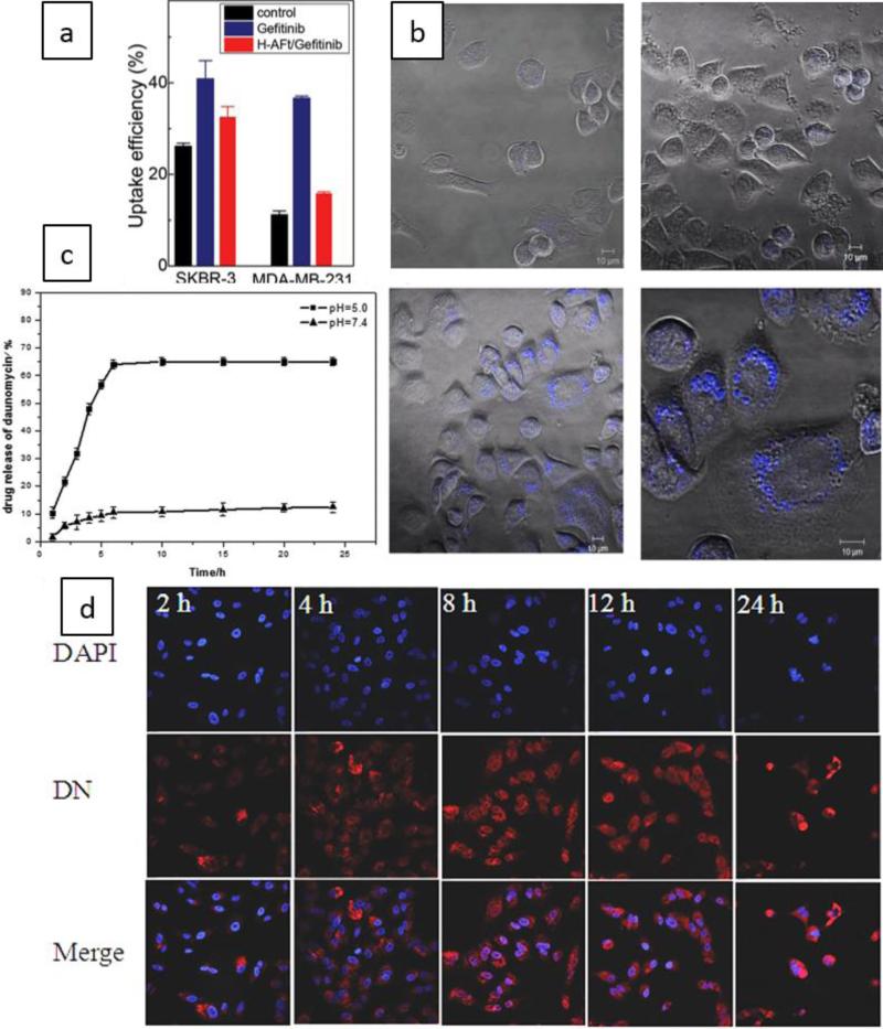
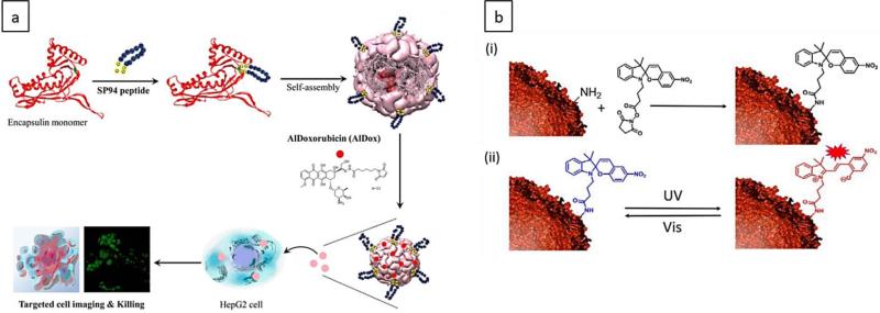
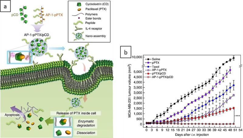
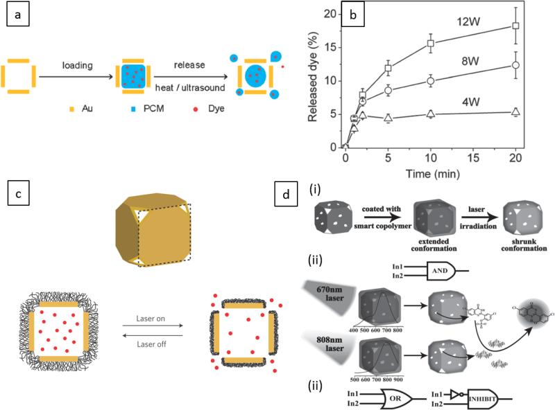
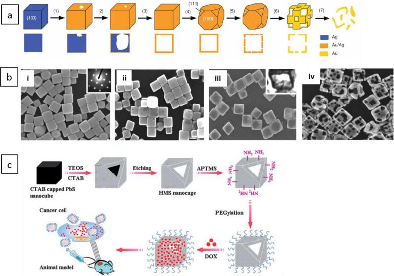
Nanocages (NCs) have emerged as a new class of drug-carriers, with a wide range of possibilities in multi-modality medical treatments and theranostics. Nanocages can overcome such limitations as high toxicity caused by anti-cancer chemotherapy or by the nanocarrier itself, due to their unique characteristics. These properties consist of: (1) a high loading-capacity (spacious interior); (2) a porous structure (analogous to openings between the bars of the cage); (3) enabling smart release (a key to unlock the cage); and (4) a low likelihood of unfavorable immune responses (the outside of the cage is safe). In this review, we cover different classes of NC structures such as virus-like particles (VLPs), protein NCs, DNA NCs, supramolecular nanosystems, hybrid metal-organic NCs, gold NCs, carbon-based NCs and silica NCs. Moreover, NC-assisted drug delivery including modification methods, drug immobilization, active targeting, and stimulus-responsive release mechanisms are discussed, highlighting the advantages, disadvantages and challenges. Finally, translation of NCs into clinical applications, and an up-to-date assessment of the nanotoxicology considerations of NCs are presented.


Figures

Figure 1
Figure 1
Schematic illustration of various nanocaged platforms, their ability to target cells and intracellular delivery via receptor-mediated uptake.
Figure 2
Figure 2
Schematic illustration presenting the wide range of capabilities and applications of caged nanoplatforms in nanomedicine and DDSs for delivery of various therapeutic drugs and imaging agents.
Figure 3
Figure 3
a) Schematic illustration of (i) riluzole loaded in iron oxide NC and (ii) blocking sodium ion channels by this NC, which prevents glutamate receptor activation and glutamate secretion, therefore reducing cancer cell growth. Reprinted with permission from ref. Copyright 2016 American Chemical Society, b) Schematic illustration of cancer treatment in mice with RBC-coated Au NCs (RBC-AuNCs). Reprinted with permission from ref. Copyright 2014 the American Chemical Society, c) Schematic of receptor-mediated cellular uptake of a biotin-functionalized truncated octahedral DNA NC by LOX-1 overexpressing cells (e.g. fibroblast cells) Reprinted with permission from ref. Copyright 2016 the American Chemical Society.
Figure 4
Figure 4
Cryo-EM image of a VLP-19 with approximately 30 nm diameter, to which palivizumab Fabs are bound through the spikes on its surface. Scale bar: 20 nm, Reprinted from ref. copyright 2015, “American Society for Clinical Investigation” (ASCI) (open access), b) Schematic illustrating a GE11 polypeptide-crosslinked MS2 VLP vector, and its uptake into cells by clathrin-mediated endocytosis for delivery of MEG3 RNA and targeting of the EGFR-positive HCC cancer cells, Reprinted from ref. Copyright 2016 “Impact Journals” (open access).
Figure 5
Figure 5
a) Presentation of a target protein onto the exterior of a P22 VLP via a decoration protein (Dec). A genetic fusion facilitated binding of the target protein to the C-terminus of the Dec (green). A poly histidine tag (purple) was retained intact on the N-terminus of Dec to facilitate purification. The formed structure was used for exterior decoration of a P22 VLP (grey). Reprinted with permission from ref. , Copyright 2015 American Chemical Society, b) Encapsulation of a CD–drug complex in VLP NCs via disulfide bonds and glutathione (GSH)-triggered release. Reprinted with permission from ref. Copyright 2013 the Royal Society of Chemistry.
Figure 6
Figure 6
a) Confocal microscopy images of incubation of PC-3 cells with viral NCs including Qβ–O488 and C60-conjugated Qβ–O488 and the subsequent cellular uptake (green). Cell nucleus and cell membrane were stained as shown in blue and red, b) cell viability using Qβ–O488 and C60-conjugated Qβ–O488 with and without phototherapy, indicating efficacy of cancer cell killing, Reprinted with permission from ref. , Copyright 2012 Royal Society of Chemistry.
Figure 7
Figure 7
Schematics illustrating (a) fabrication of a ferritin NC, followed by encapsulation of cargo molecules in its cavity through a reversible disassembly/reassembly process. This NC could be disassembled into its subunits at pH equal to 4.0, and then reassembled into the protein NC form at pH=7.5, b) apoferritin NC disassembled to its subunits at pH=11.0, followed by encapsulation of cargo molecules (lutein) and NC assembly at pH=6.0. Chitosan was used to form a polyelectrolyte complex of FCLs via electrostatic interactions with exterior surface of the apoferritin. a and b Reprinted with permission from ref. and Copyright 2016, Royal Society of Chemistry.
Figure 8
Figure 8
a) Mean uptake efficiency by SKBR3 and MDA-MB-231 cells obtained from flow cytometry, b) Images of confocal microscopy indicating cellular uptake of control sample (upper images) and Gefitinib-loaded apoferritin NCs by SKBR3 cells (lower images) , Open Access, John Wiley & Sons, Inc., c) The curve indicating enhanced drug release for a DN-loaded pH-sensitive HA surface-modified apoferritin in more acidic conditions, d) Confocal images showing Internalization of the nanocarrier into A549 cells at different times (2, 4, 8, 12, and 24 hour), Reprinted with permission from ref. , Copyright 2015 Royal Society of Chemistry.
Figure 9
Figure 9
a) Encapsulin NCs for targeting the surface markers of HCC cells using SP94-peptide (blue) with linker (yellow) genetically or chemically attached (green) to the exterior surface of the NC. AlDOX is chemically attached to Encap_loophis42C123 and delivered to the target cell. Reprinted with permission from ref. Copyright 2014 the American Chemical Society. b) Schematic showing integration of a light-responsive moiety (spiropyran) with an encapsulin NC at its surface through amine-succinimide reaction (i), and UV light-activated isomerization of spiropyran moiety (blue) with no fluorescence to merocyanine moiety (red) with high fluorescent activity. This reaction is reversible via a visible light irradiation (ii) Reprinted with permission from ref. Copyright 2016, John Wiley & Sons, Inc.
Figure 10
Figure 10
a) Conjugation of CD and PTX molecules via their hydroxyl groups to copolymer backbone led to enzyme degradation of the ester linkages hydrolysis inducing apoptosis in cancer cells . b) Growth profile of MDA-MB-231 tumors, respectively treated with different groups. (pPTX / pCD: polymeric PTX/ polymeric CD. AP-1: peptide ligand). Reproduced with permission from ref. Copyright 2014 Nature Publishing Group.
Figure 11
Figure 11
a) Schematic illustration of loading the hollow interior of AuNCs with a drug-doped PCM and its release from the AuNCs by direct or ultrasonic heating. b) Release profiles of the dye exposed to HIFU at different applied powers and time points. Reprinted with permission from ref. Copyright 2011 the American Chemical Society, c) photothermal triggered release of encapsulated cargos from PNIPAAm-conjugated AuNCs due to NIR-irradiation induced temperature increase. This allowed LCST transition of PNIPAAm moieties, leading to their hydrophobicity and shrinkage, which induced release of the cargos, d) logic-gated operation of polymer shell conjugated AuNCs , which can perform various logic-gates including AND, OR, and INHIBIT and lead to logic-gated cargo release. Reprinted with permission from ref. , Copyright 2014 John Wiley & Sons, Inc.
Figure 12
Figure 12
a) Schematic illustration of the major steps of the production of AuNCs from Ag nanocubes by the galvanic replacement method. (1) Starting reaction; (2) Production of initial hollow nanostructure; (3) Formation of nanoboxes; (4) Dealloying of the Au/Ag nanoboxes; (5) Formation of AuNC-nanobox with pores in the walls; (6) Production of AuNCs. Reprinted with permission from ref. Copyright 2007 Nature Publishing group, b) SEM images (i, ii, iii and iv) (E) of morphological changes in formation of Au NCs: starting from conversion of Ag nanocubes into Au/Ag nanoboxes, and finaly into Au NCs. Reprinted with permission from ref. Copyright 2004 the American Chemical Society, c) Schematic showing synthesis of hollow mesoporous silica NCs and their modification for drug delivery. [CTAB: cetyltrimethylammonium bromide; TEOS: tetraethyl orthosilicate; APTMS: 3-aminopropyltriethoxysilane; PEG: polyethylene glycol; DOX: doxorubicin]. Reprinted with permission from ref. Copyright 2011 the Royal Society of Chemistry.

Similar articles

Cited by

References

    1. Sahandi Zangabad P, Khodabakhshi F, Simchi A, Kokabi AH. International Journal of Fatigue. 2016;87:266–278.
    1. Mahmoudi N, Simchi A. Materials Science and Engineering: C. 2017;70:121–131. - PubMed
    1. Mahdi Karimi HZ, Sahandi Zangabad Parham, Bakhshian Nik Amirala, Yazdani Narges, Hamrang Mohammad, Mohamed Elmira, Masoud Moosavi Basri Seyed, Bakhtiari Leila, Hamblin MR. Nanomedicine, Future Medicine. 2016 - PMC - PubMed
    1. Huang Y-Y, Choi H, Kushida Y, Bhayana B, Wang Y, Hamblin MR. Antimicrobial Agents and Chemotherapy. 2016;60:5445–5453. - PMC - PubMed
    1. Mirkin CA, Meade TJ, Petrosko SH, Stegh AH. Nanotechnology-Based Precision Tools for the Detection and Treatment of Cancer. Springer; 2015.

LinkOut - more resources