. 2017 Feb 1;127(2):543-548.
doi: 10.1172/JCI89364. Epub 2017 Jan 9.

Versatile humanized niche model enables study of normal and malignant human hematopoiesis

Versatile humanized niche model enables study of normal and malignant human hematopoiesis

Ander Abarrategi et al. J Clin Invest. .

Abstract

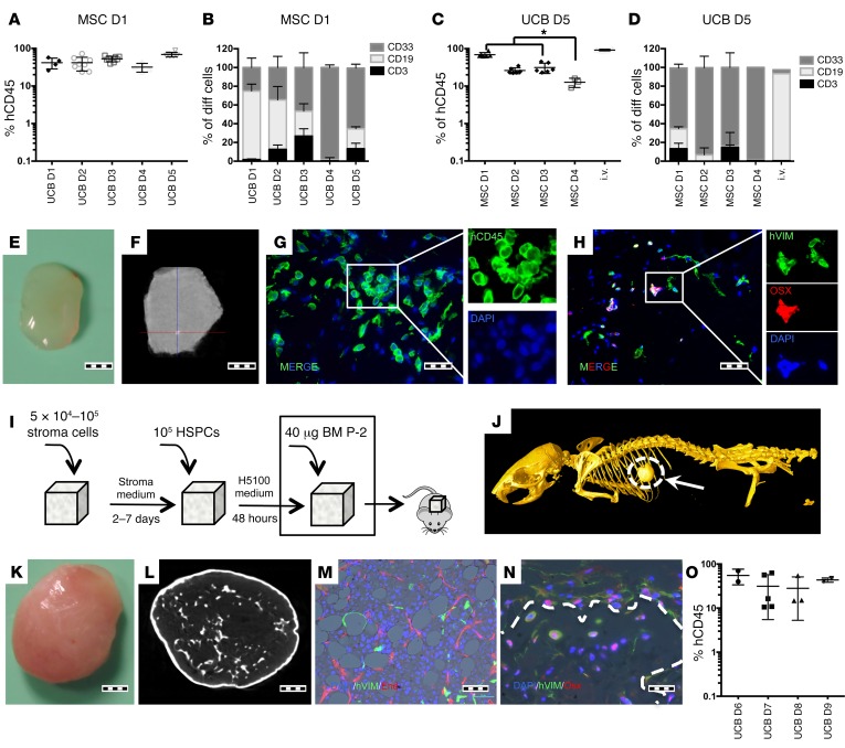
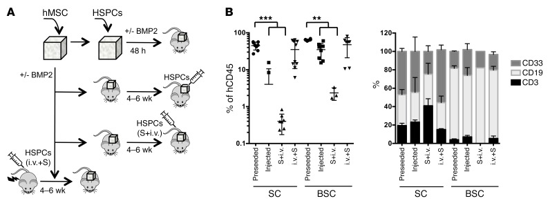
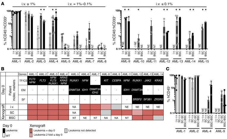
The BM niche comprises a tightly controlled microenvironment formed by specific tissue and cells that regulates the behavior of hematopoietic stem cells (HSCs). Here, we have provided a 3D model that is tunable in different BM niche components and useful, both in vitro and in vivo, for studying the maintenance of normal and malignant hematopoiesis. Using scaffolds, we tested the capacity of different stromal cell types to support human HSCs. Scaffolds coated with human mesenchymal stromal cells (hMSCs) proved to be superior in terms of HSC engraftment and long-term maintenance when implanted in vivo. Moreover, we found that hMSC-coated scaffolds can be modulated to form humanized bone tissue, which was also able to support human HSC engraftment. Importantly, hMSC-coated humanized scaffolds were able to support the growth of leukemia patient cells in vivo, including the growth of samples that would not engraft the BM of immunodeficient mice. These results demonstrate that an s.c. implantation approach in a 3D carrier scaffold seeded with stromal cells is an effective in vivo niche model for studying human hematopoiesis. The various niche components of this model can be changed depending on the context to improve the engraftment of nonengrafting acute myeloid leukemia (AML) samples.


Conflict of interest statement

The authors have declared that no conflict of interest exists.

Figures

Figure 1
Figure 1. UCB-derived HSPC engraftment in s.c. scaffolds.
(A and B) Comparative assay using a single hMSC donor and multiple UCB donors. Each point represents 1 mouse seeded with 6 scaffolds. For each UCB donor, 3 to 8 mice were transplanted. (A) hCD45 scaffold-engraftment level. D1-D5: different UCB Donors. (B) Lineage distribution of engrafted cells. (C and D) A comparative assay using a single UCB donor and multiple hMSC donors. Each point represents 1 mouse seeded with 6 scaffolds. For each hMSC donor, 3 to 6 mice were transplanted. (C) hCD45 scaffold–engraftment levels. For comparative purposes, the engraftment levels in BM of i.v.-injected mice are provided. Tukey’s test for multiple comparisons was applied. *P < 0.05. (D) Lineage distribution of engrafted cells. (E) Gross morphology of harvested scaffold. (F) microCT study of a harvested scaffold. The scaffold is mainly soft tissue, with sporadic calcification spots. (G) Immunofluorescence image showing hCD45 cells in the scaffold and (H) osterix-positive (OSX+) and osterix-negative human stroma cells. (I) Schematic of preparation and implantation of bone-forming scaffolds. (J) Whole-mouse microCT showing the s.c. ossicle formation. (K) Gross morphology of the harvested ossicle. (L) microCT study of a harvested ossicle. (M and N) Immunofluorescence images showing (M) human vimentin+ (hVIM) mesenchymal cells and adipocytes and mature neovascularized (endomucin+, End) BM. (N) Trabecular bone formed inside ossicles. Dotted line shows delimited trabecular bone area. hVIM+/Osx+ cells are osteocytes or osteoblasts in the bone surface areas. (O) hCD45+ engraftment levels in the ossicle model tested with multiple UCB donors. Each point represents 1 mouse with 2 scaffolds. For each UCB donor, 2 to 4 mice were transplanted. Scale bars: 1 mm (E, F, K, and L); 20 μm (G, H and N); 50 μm (M). All data were harvested at 12 weeks after implantation.
Figure 2
Figure 2. Different modality of transplantation of HSPCs into scaffold.
(A) Schematic of hMSC seeding and the different tested methods for CB-HSPCs inclusion in the scaffold: preseeded in the scaffolds before implantation in nonconditioned mice (preseeded); intrascaffold injection of hHSPCs in nonconditioned mice; i.v. injection of hHSPCs in scaffold-implanted nonconditioned mice (S+i.v.); and finally, irradiated mice injected i.v. with hHSPCs and 4 to 6 weeks later implanted with hMSC-seeded scaffolds (i.v.+S). All assays were performed without BMP2 (SC) or with BMP2 injection (BSC). All assays were performed with pools of HSPCs from different donors. (B) Analysis of human cells in the scaffolds. Percentage of hCD45+ cells (left panel) and proportion of hCD3+, hCD19+, and hCD33+ cells in the different scaffolds 12 weeks after implantation of HSPCs. Each point represents 1 mouse implanted with 2 to 6 scaffolds, which were pooled before the analysis. For each condition, 3 to 10 mice were transplanted. Preseeded approach shows higher engraftment levels than injected and S+i.v. approaches. Tukey’s multiple comparison test was applied: **P ≤ 0.005; ***P ≤ 0.0005.
Figure 3
Figure 3. AML engraftment in different implantation models.
(A) Percentage of hCD45+hCD33+ cells recovered from each xenograft implantation model compared with conventional i.v. injection in NSG mice 12 weeks after transplant for patients classified as high engrafters (≥ 1% engraftment), low engrafters (between 1 to 0.1%), and nonengrafters (< 0.1%) using conventional i.v. injection. i.v., i.v. injection; SC, scaffold-implantation; BSC, bone scaffold-implantation. Comparison between i.v. and SC or BSC was applied using Dunnett’s test. *P < 0.05. Each point represents 1 mouse implanted with 2 to 6 scaffolds. For each patient, 2 to 8 mice were transplanted per condition. (B) Graphical representation of gene mutations detected in CD33+ pretransplant and hCD45+hCD33+ postxenografted cells. Mutations are grouped in transcription factor and cell signaling (TF/CS) genes, epigenetic modifier (EM) genes, and splicing factor (SF) genes. NA, sample not available for testing; NE, no engraftment was detected in mice; NT, not tested. Red boxes, variant allele frequencies comparable to day 0; pink boxes, ≤ 2-fold day 0; gray boxes, not detected. Variant allele frequencies for xenotransplanted samples are the average between ≥1, where applicable. (C) Percentage of hCD45+ cells recovered from secondary recipients. Primary cells from i.v. mice were transplanted i.v. into secondary mice (i.v.-i.v.). Primary cells from SC were transplanted i.v. (SC-i.v.) or implanted into scaffold (SC-SC) in secondary mice. Primary cells from BSC were implanted into BSC in secondary mice (BSC-BSC). Each point represents 1 mouse. For each patient, 2 to 4 mice were transplanted per condition.

References

    1. Pearce DJ, et al. AML engraftment in the NOD/SCID assay reflects the outcome ofAML: implications for our understanding of the heterogeneity of AML. Blood. 2006;107(3):1166–1173. - PMC - PubMed
    1. Vargaftig J, et al. Frequency of leukemic initiating cells does not depend on the xenotransplantation model used. Leukemia. 2012;26(4):858–860. - PMC - PubMed
    1. Sanchez PV, et al. A robust xenotransplantation model for acute myeloid leukemia. Leukemia. 2009;23(11):2109–2117. - PMC - PubMed
    1. Theocharides AP, Rongvaux A, Fritsch K, Flavell RA, Manz MG. Humanized hemato-lymphoid system mice. Haematologica. 2016;101(1):5–19. doi: 10.3324/haematol.2014.115212. - DOI - PMC - PubMed
    1. Lee J, et al. Implantable microenvironments to attract hematopoietic stem/cancer cells. Proc Natl Acad Sci USA. 2012;109(48):19638–19643. doi: 10.1073/pnas.1208384109. - DOI - PMC - PubMed

MeSH terms