. 2017 Feb 1;127(2):564-568.
doi: 10.1172/JCI90520. Epub 2017 Jan 9.

MerTK receptor cleavage promotes plaque necrosis and defective resolution in atherosclerosis

MerTK receptor cleavage promotes plaque necrosis and defective resolution in atherosclerosis

Bishuang Cai et al. J Clin Invest. .

Abstract

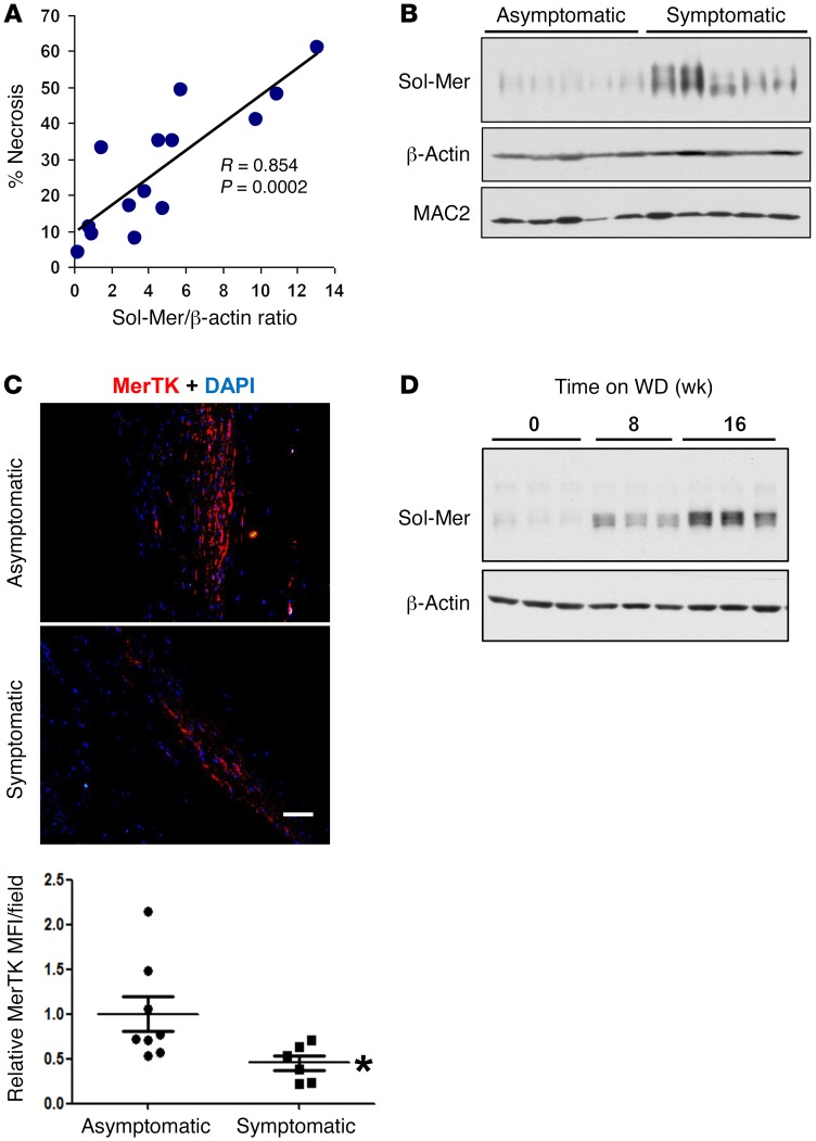
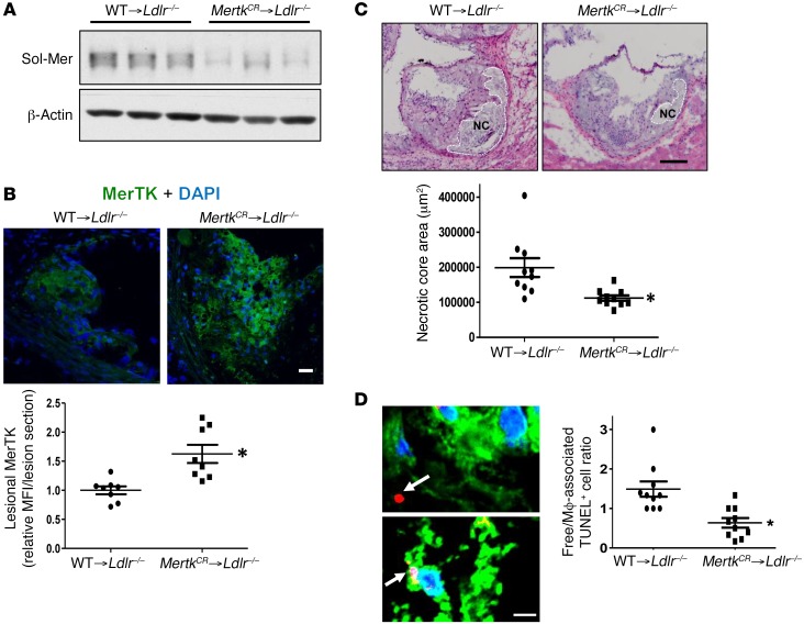
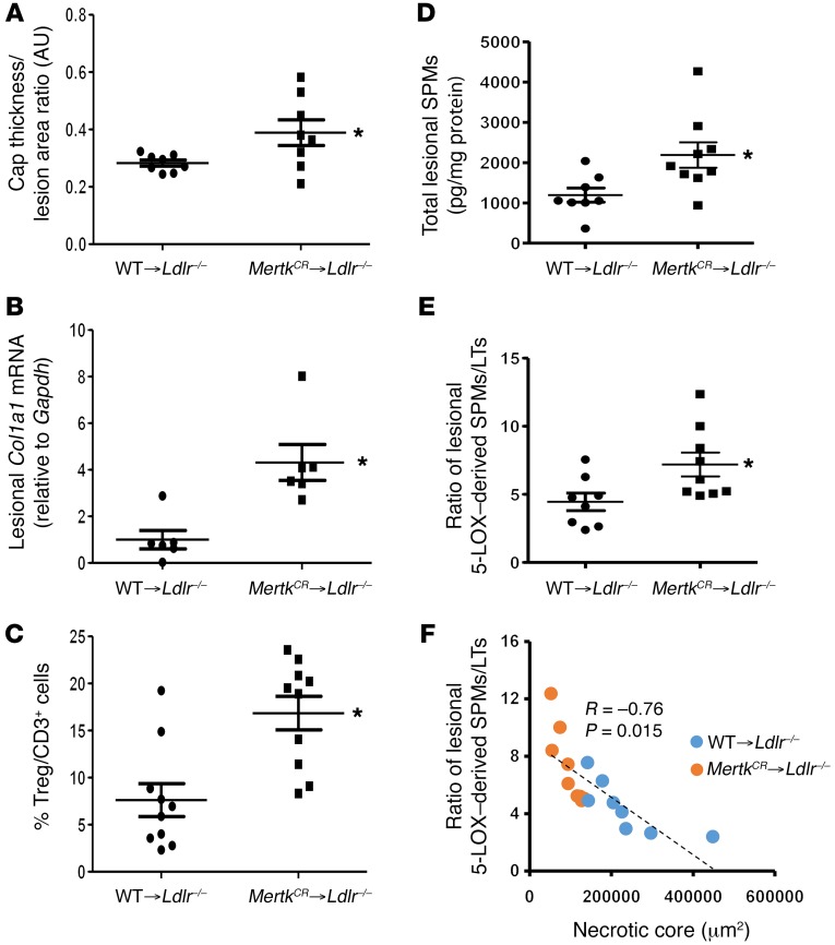
Atherothrombotic vascular disease is often triggered by a distinct type of atherosclerotic lesion that displays features of impaired inflammation resolution, notably a necrotic core and thinning of a protective fibrous cap that overlies the core. A key cause of plaque necrosis is defective clearance of apoptotic cells, or efferocytosis, by lesional macrophages, but the mechanisms underlying defective efferocytosis and its possible links to impaired resolution in atherosclerosis are incompletely understood. Here, we provide evidence that proteolytic cleavage of the macrophage efferocytosis receptor c-Mer tyrosine kinase (MerTK) reduces efferocytosis and promotes plaque necrosis and defective resolution. In human carotid plaques, MerTK cleavage correlated with plaque necrosis and the presence of ischemic symptoms. Moreover, in fat-fed LDL receptor-deficient (Ldlr-/-) mice whose myeloid cells expressed a cleavage-resistant variant of MerTK, atherosclerotic lesions exhibited higher macrophage MerTK, lower levels of the cleavage product soluble Mer, improved efferocytosis, smaller necrotic cores, thicker fibrous caps, and increased ratio of proresolving versus proinflammatory lipid mediators. These findings provide a plausible molecular-cellular mechanism that contributes to defective efferocytosis, plaque necrosis, and impaired resolution during the progression of atherosclerosis.


Conflict of interest statement

The authors have declared that no conflict of interest exists.

Figures

Figure 1
Figure 1. Sol-Mer is increased in advanced human and murine atherosclerotic lesions.
(A) Carotid endarterectomy (CEA) specimens were analyzed for sol-Mer by immunoblot, and necrotic area was measured from H&E-stained images. The correlation coefficient (R) and P value were based on Pearson’s product-moment correlation analysis. (B) CEA specimens from asymptomatic and symptomatic patients were assessed by immunoblot for sol-Mer (n = 5 per group). β-Actin and MAC2 were used as loading controls for total protein and macrophages, respectively. (C) Paraffin-embedded CEA sections were stained with anti-MerTK antibody, imaged by confocal microscopy (scale bar: 30 μm), and quantified by MerTK mean fluorescence intensity (MFI) relative to the asymptomatic cohort (n = 6–8 for each group). *P < 0.05, by 2-tailed Student’s t test. (D) Age-matched Ldlr–/– mice were fed the Western-type diet (WD) for 0, 8, or 16 weeks and sacrificed at 24 weeks of age. The aortic arch, Brachiocephalic Artery (BCA), and descending aorta up to the renal bifurcation were removed en bloc and immunoblotted for sol-Mer (n = 3 per group).
Figure 2
Figure 2. Suppression of MerTK cleavage in myeloid cells improves efferocytosis and decreases plaque necrosis in WD-fed Ldlr–/– mice.
(A) WT → Ldlr–/– or MertkCRLdlr–/– bone marrow–transplanted mice were fed WD for 16 weeks. Aortas were harvested and analyzed for sol-Mer as described in Figure 1D (n = 3 per group). *P < 0.05. (B) Confocal microscopy of MerTK in aortic root cross sections (scale bar: 20 μm). Images were quantified as MerTK MFI relative to the WT → Ldlr–/– cohort (n = 8 per group). *P < 0.01. (C) Representative H&E images of aortic root sections, with necrotic core (NC) regions indicated by broken lines (scale bar: 40 μm), and quantification of necrotic core area (n = 10 per group, 2 independent experiments). *P < 0.01. (D) Representative images of aortic root sections in which apoptotic cells were labeled by TUNEL (red), macrophages by anti-F4/80 (green), and nuclei by Hoechst (blue) (scale bar: 10 μm). The white arrows depict apoptotic cells that were either free (top image) or associated with macrophages (bottom image). The graph shows quantification of the ratio of free to macrophage-associated apoptotic cells (n = 10 for each group, 2 independent experiments). *P < 0.05. A 2-tailed Student’s t test was used for all panels.
Figure 3
Figure 3. Suppression of MerTK cleavage improves features of resolution in plaques and increases aortic content of specialized proresolving mediators.
(A) Aortic root sections from WT → Ldlr–/– and MertkCRLdlr–/– bone marrow–transplanted mice were stained with Picrosirius red. Quantified data are presented as the ratio of fibrous cap thickness to lesion area, expressed as arbitrary units (AU) (n = 8 per group). *P < 0.05. (B) A subset of aortic root sections was chosen randomly for quantification of Col1a1 mRNA by reverse transcriptase quantitative PCR, with normalization to Gapdh mRNA (n = 6 for each group). *P < 0.05. (C) FoxP3+ Tregs and total CD3+ T cells were quantified in aortic root sections by immunofluorescence microscopy and expressed as percentage Tregs/CD3+ cells (n = 10 for each group). The average absolute numbers of these cells, most of which were in the adventitia, were 8 ± 2.49 and 19 ± 3.74 per section for FoxP3+ Tregs (*P < 0.05) and 106 ± 16.13 and 107 ± 14.33 per section for CD3+ cells (NS) for the WT and MertkCR cohorts, respectively. (D and E) Quantification of specialized proresolving mediators (SPMs) and ratio of aortic 5-LOX–derived SPMs/leukotrienes (LTs) in aorta of WT → Ldlr–/– (n = 8) versus MertkCRLdlr–/– (n = 9) bone marrow–transplanted mice. *P < 0.05. (F) Correlation of the ratio of 5-LOX–derived SPMs/leukotrienes with necrotic core area (n = 17). P represents the 2-tailed probability value of a Pearson correlation coefficient. A 2-tailed Student’s t test was used for AE.

References

    1. Virmani R, Kolodgie FD, Burke AP, Farb A, Schwartz SM. Lessons from sudden coronary death: a comprehensive morphological classification scheme for atherosclerotic lesions. Arterioscler Thromb Vasc Biol. 2000;20(5):1262–1275. doi: 10.1161/01.ATV.20.5.1262. - DOI - PubMed
    1. Schrijvers DM, De Meyer GR, Kockx MM, Herman AG, Martinet W. Phagocytosis of apoptotic cells by macrophages is impaired in atherosclerosis. Arterioscler Thromb Vasc Biol. 2005;25(6):1256–1261. doi: 10.1161/01.ATV.0000166517.18801.a7. - DOI - PubMed
    1. Tabas I. Consequences and therapeutic implications of macrophage apoptosis in atherosclerosis: the importance of lesion stage and phagocytic efficiency. Arterioscler Thromb Vasc Biol. 2005;25(11):2255–2264. doi: 10.1161/01.ATV.0000184783.04864.9f. - DOI - PubMed
    1. Merched AJ, Ko K, Gotlinger KH, Serhan CN, Chan L. Atherosclerosis: evidence for impairment of resolution of vascular inflammation governed by specific lipid mediators. FASEB J. 2008;22(10):3595–3606. doi: 10.1096/fj.08-112201. - DOI - PMC - PubMed
    1. Tabas I. Macrophage death and defective inflammation resolution in atherosclerosis. Nat Rev Immunol. 2010;10(1):36–46. - PMC - PubMed

MeSH terms