Observational Study
. 2017 Oct;37(10):1905-1915.
doi: 10.1097/IAE.0000000000001443.

ACUTE CENTRAL SEROUS CHORIORETINOPATHY: Factors Influencing Episode Duration

Affiliations
Observational Study

ACUTE CENTRAL SEROUS CHORIORETINOPATHY: Factors Influencing Episode Duration

Alejandra Daruich et al. Retina. 2017 Oct.

Abstract

Purpose: To evaluate the influence of clinical and multimodal imaging parameters on the duration of acute central serous chorioretinopathy (CSCR) episodes.

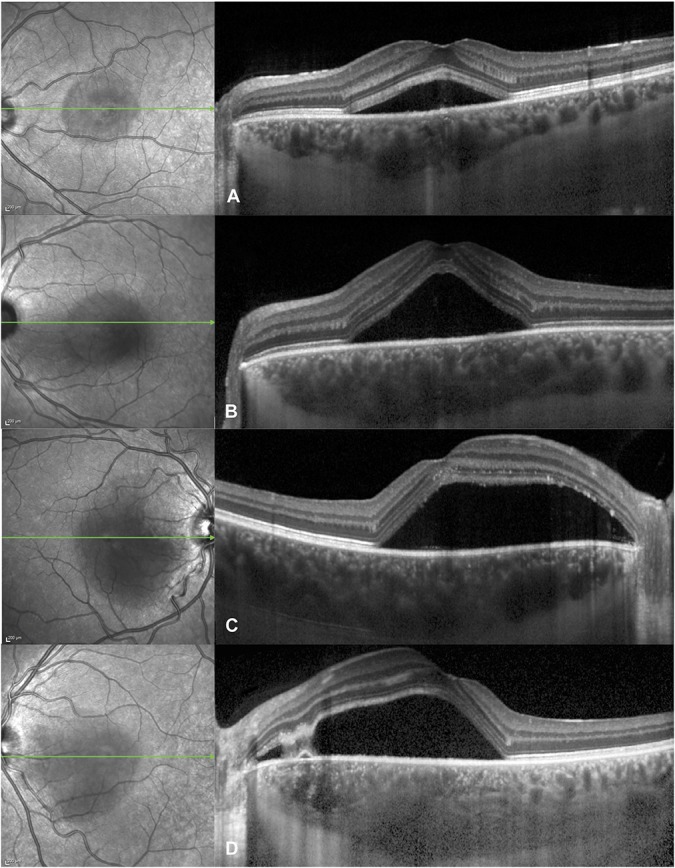
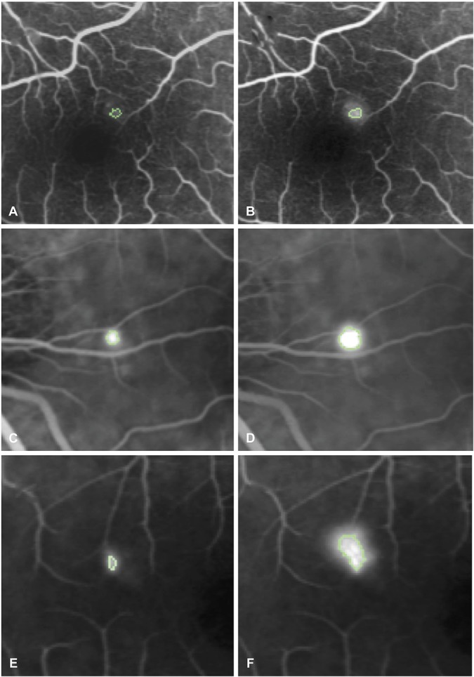
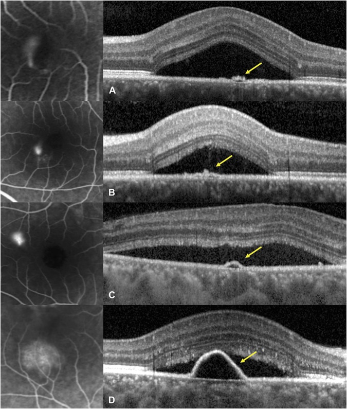
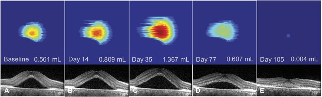
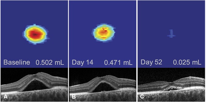
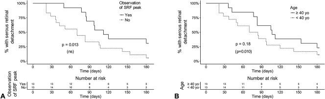
Methods: Consecutive patients with first, treatment-naïve central serous chorioretinopathy episodes presenting within 20 days of symptoms onset were prospectively included. They were reevaluated 15 days to 20 days later, followed by monthly evaluation for 6 months. Subfoveal choroidal thickness (SFCT), fluorescein leakage intensity on fluorescein angiography, elevation of retinal pigment epithelium (RPE) lesions at leakage sites, focal/multifocal pattern of indocyanine green angiography (ICGA) at baseline, time-dependent pattern of subretinal fluid (SRF) resorption on OCT using volume segmentation, history of corticosteroid intake and mean blood pressure were evaluated using univariate (Log rank test) and multivariate (Cox proportional hazard regression) survival analysis.

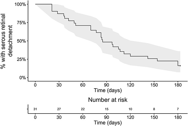
Results: Thirty-one patients were included (26 men, 5 women, mean age: 40.0 ± 8.9 years, range: 24-58), of which 26 (84%) had episode resolution by 6 months. Using univariate analysis, episode duration was longer in cases with subfoveal choroidal thickness ≥500 μm (P = 0.0002), retinal pigment epithelium elevation at leakage sites ≥50 μm (P = 0.033), and a peak in subretinal fluid observed during follow-up (P = 0.013), and there was a near-significant association of intense fluorescein leakage (P = 0.074) with longer episodes. Using multivariate analysis, subfoveal choroidal thickness ≥500 μm (P = 0.017), retinal pigment epithelium elevation at leakage sites ≥50 μm (P = 0.010) and patient age ≥40 years (P = 0.010) were significantly and independently associated to longer episodes. Indocyanine green angiography pattern, corticosteroid intake, and blood pressure did not influence episode duration.

Conclusion: Older age, higher subfoveal choroidal thickness, and higher degree of retinal pigment epithelium alteration at leakage sites are independent factors of longer acute central serous chorioretinopathy episodes.


Conflict of interest statement

None of the authors has any financial/conflicting interests to disclose.

Figures

Fig. 1.
Fig. 1.
Survival curve showing the time-dependent resolution rate of 31 acute episodes of central serous chorioretinopathy from the time of the initial visit. All patients presented within 20 days after onset of symptoms. By 6 months, resolution was observed in 26 patients. The 95% confidence interval is colored in gray.
Fig. 2.
Fig. 2.
Spectrum of subfoveal choroidal thickness in acute central serous chorioretinopathy, visible on optical coherence tomography scans (right) passing through the fovea as indicated by the green arrows on infrared images (left): (A) 403 mm in a 37-year-old man, (B) 469 mm in a 28-year-old man, (C) 519 mm in a 38-year-old man, and (D) 617 mm in a 34-year-old-man.
Fig. 3.
Fig. 3.
Method to quantify fluorescein leakage expansion on fluorescein angiography from early phase (40–60 seconds) to midphase (2–2.5 minutes). A and B. Early phase (A) and midphase (B) angiograms in a case with a very weak leakage. The hyperfluorescent area corresponding to pixels whose intensity is comprised within 75% of the maximal hyperfluorescence is indicated by a green outline. The fluorescein expansion ratio was calculated as the ratio between the hyperfluorescent areas at midphase and early phase and was 1.32. C and D. Same method applied to early phase (C) and midphase (D) angiograms from a case with intermediate ink-blot leakage pattern, yielding a fluorescein expansion ratio of 3.98. E and F. Same method applied to early (E) and midphase (F) angiograms from a case with intense smokestack leakage pattern, yielding a fluorescein expansion ratio of 6.01.
Fig. 4.
Fig. 4.
Optical coherence tomography of pigment epithelial lesions (right) at leaking sites on fluorescein angiograms (left) in acute central serous chorioretinopathy. A and B. Pigment epithelial bumps (yellow arrows) with estimated height of 25 mm in a 36-year-old man (A) and 41mm in a 28-year-old man (B). C and D. Pigment epithelial detachments (yellow arrows) with estimated height of 54 mm in a 39-year-old man(C) and 163 mm in a 52-year-old woman (D).
Fig. 5.
Fig. 5.
Follow-up of an acute episode of central serous chorioretinopathy in a 52-year-old woman using subretinal fluid volume segmentation on optical coherence tomography. There was an initial increase in subretinal fluid from baseline (AB) with a peak in subretinal fluid volume at Day 35 (C) and subsequent decrease (DE) until subretinal fluid resolution at Day 132 (not shown). Note the shape of a pigment epithelial detachment visible on the segmented serous retinal detachment (same patient as Figure 4D).
Fig. 6.
Fig. 6.
Follow-up of an acute episode of central serous chorioretinopathy in a 35-year-old man using subretinal fluid volume segmentation on optical coherence tomography. No increase in subretinal fluid volume could be observed, with a progressive decrease from baseline (A) to all timepoints (B and C) and resolution at Day 75 (not shown).
Fig. 7.
Fig. 7.
Comparative survival curves showing the rate of subretinal detachment resolution over time about SFCT (A) and the height of RPE elevations at leakage sites (B) on optical coherence tomography. The duration from initial visit to episode resolution was significantly longer for subfoveal choroidal thickness ≥500 μm (A) and pigment epithelium elevation ≥50 μm (B) both in the univariate and multivariate analyses. P-values from the univariate (log-rank test) and the multivariate analysis (Cox proportional hazard model, between parenthesis) are reported.
Fig. 8.
Fig. 8.
Comparative survival curves showing the rate of subretinal detachment resolution over time about the observation of a peak in SRF on optical coherence tomography (A) and patient age (B). The duration from initial visit to episode resolution was significantly longer for cases with an observed peak in subretinal fluid volume in the univariate analysis only (A). Episodes were also longer for patients aged 40 years or older in the multivariate analysis only (B). P-values from the univariate (log-rank test) and the multivariate analysis (Cox proportional hazard model, between parenthesis) are reported. ns, nonsignificant contributor to the multivariate model.

References

    1. Klein ML, Van Buskirk EM, Friedman E, et al. Experience with nontreatment of central serous choroidopathy. Arch Ophthalmol 1974;91:247–250. - PubMed
    1. Leaver P, Williams C. Argon laser photocoagulation in the treatment of central serous retinopathy. Br J Ophthalmol 1979;63:674–677. - PMC - PubMed
    1. Robertson DM, Ilstrup D. Direct, indirect, and sham laser photocoagulation in the management of central serous chorioretinopathy. Am J Ophthalmol 1983;95:457–466. - PubMed
    1. Daruich A, Matet A, Dirani A, et al. Central serous chorioretinopathy: recent findings and new physiopathology hypothesis. Prog Retin Eye Res 2015;48:82–118. - PubMed
    1. Malik KJ, Sampat KM, Mansouri A, et al. Low-intensity/high-density subthreshold micropulse diode laser for chronic central serous chorioretinopathy. Retina 2015;35:532–536. - PubMed

Publication types