Review
. 2017 Jan 5;6(1):1.
doi: 10.3390/biology6010001.

Mechanisms to Avoid and Correct Erroneous Kinetochore-Microtubule Attachments

Affiliations
Review

Mechanisms to Avoid and Correct Erroneous Kinetochore-Microtubule Attachments

Michael A Lampson et al. Biology (Basel). .

Abstract

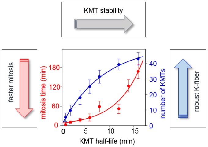
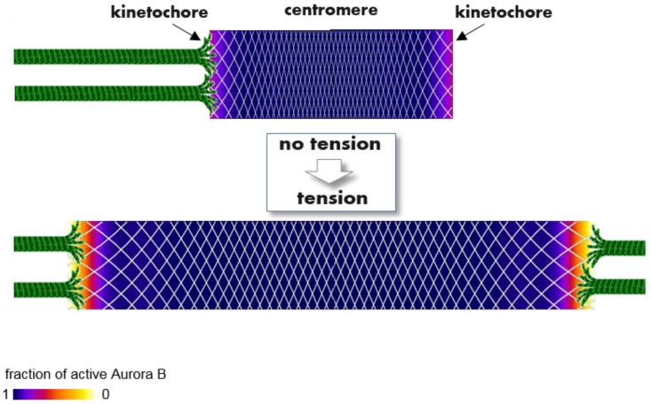
In dividing vertebrate cells multiple microtubules must connect to mitotic kinetochores in a highly stereotypical manner, with each sister kinetochore forming microtubule attachments to only one spindle pole. The exact sequence of events by which this goal is achieved varies considerably from cell to cell because of the variable locations of kinetochores and spindle poles, and randomness of initial microtubule attachments. These chance encounters with the kinetochores nonetheless ultimately lead to the desired outcome with high fidelity and in a limited time frame, providing one of the most startling examples of biological self-organization. This chapter discusses mechanisms that contribute to accurate chromosome segregation by helping dividing cells to avoid and resolve improper microtubule attachments.

Keywords: Aurora B kinase; kinetochore geometry; microtubule turnover; tension-dependent regulation.


Conflict of interest statement

The authors declare no conflict of interest.

Figures

Figure 1
Figure 1
Tension-dependent error correction for syntelic kinetochore microtubule (KMT) attachments. Kinetochores (red) of a meiotic bivalent (blue) repeatedly bind MTs from different poles (green), but KMT attachments in the syntelic configuration are short-lived. Tension arises when the amphitelic configuration is encountered, inducing stable KMT attachments. Syntelic attachments can be stabilized artificially by applying tension with a microneedle. In meiosis I, as shown here, kinetochores of homologous chromosomes attach to opposite spindle poles in the amphitelic orientation. In mitotic cells, kinetochores of sister chromatids would attach to opposite poles.
Figure 2
Figure 2
Merotelic KMT configuration for a congressed chromosome. Top image is a three-dimensional representation of a mammalian chromosome (green) positioned midway between two spindle poles. Sister chromatids (green) are connected by a stretchable centromere, represented as a spring (yellow). Bottom image is an enlargement of sister kinetochores, depicted as semi-transparent layers. Most of the attached MTs are in the proper amphitelic configuration (grey), extending from opposite spindle poles. However, although the chromosome has congressed and its sister kinetochores are positioned back-to-back, they can still bind improper merotelic MTs (one such MT is shown in orange). Computer-generated images are snapshots from video material in [23].
Figure 3
Figure 3
Models for correction of merotelic KMT attachment errors. Correction of merotelic attachments in vertebrate cells proceeds gradually during prometaphase and metaphase without necessarily losing all KMT attachments. (A) In the tension-dependent error-correction model, stretching between sister kinetochores (red ovals connected with blue springs) is the primary signal that modulates the turnover of KMT attachments (depicted with curved arrows). As kinetochores bind and lose KMTs, probing different configurations, the configurations that produce higher tension are assumed to induce higher KMT stability. Because their KMTs detach less frequently, such configurations last longer. This gradual evolution would lead eventually to the completely amphitelic configuration, which generates maximal tension. In the non-discriminate version of this model (as shown), all KMTs are affected similarly. Alternatively, in the selective version of this model, lifetime of merotelic vs. amphitelic KMTs is assumed to be regulated differently (see text for details). (B) In the basic mechanism, the gradual correction of merotelic attachments proceeds with no change in the turnover of KMT attachments. The rate of KMT turnover in this model does not depend on tension and is not selective, so all old KMTs (orange) eventually detach, whether correct and incorrect, and are replaced with new KMTs (grey). New KMTs preferentially attach correctly, favored by the back-to-back geometry of sister kinetochores.
Figure 4
Figure 4
Quantitative analysis of the evolution of KMT configurations during mitotic progression. Graphs illustrate changes in the number of merotelic KMTs during mitotic progression, as predicted by the basic error-correction model [23]. After mitosis starts, the number of attached MTs, some of which are merotelic, increases sharply. As chromosomes become aligned midway on the spindle, the number of merotelic KMTs begins to decline. However, merotelic KMTs are not eliminated completely by this mechanism because new erroneous attachments continue to form even on congressed chromosomes, albeit at lower frequency. Thus, the system tends toward a steady state, in which the rates of forming wrong attachments and eliminating them through non-discriminatory turnover are balanced. (A) With more stringent geometric constraints, the final outcome for the KMT configuration at anaphase onset is improved because the rate of capturing wrong KMTs at steady-state is reduced. (B) The rate of KMT turnover does not affect the final outcome (black arrow). However, if anaphase starts before the steady state is reached, cells with slower KMT turnover will have more merotelic KMTs.
Figure 5
Figure 5
Competing constraints on KMT turnover during mitotic progression. Graph illustrates how KMT turnover (represented as KMT half-life, horizontal axis) affects the overall speed of mitosis and the total number of acquired KMTs (based on the error-correction model in [23]). The speed of mitotic progression (left axis) is evaluated based on the time required to achieve the steady-state KMT configuration. At steady state, the number of attached KMTs (right axis) and the fraction of merotelic KMTs (not shown) have stopped changing, and increasing the duration of mitosis does not generate more KMTs or improve accuracy. When KMT turnover is slow, i.e., KMT half-life is longer, attachments are more stable because they have low release rate. The time required to release all old KMTs increases quickly with increasing KMT stability (red curve), so shorter KMT lifetime is required for speedy mitotic progression. However, the number of KMTs in a K-fiber is lower for shorter KMT lifetime (blue curve). Thus, the limited time of mitotic progression and the acquisition of a full set of KMTs represent the competing constraints on the rate of KMT turnover during mitosis.
Figure 6
Figure 6
A molecular model to explain how tension regulates phosphorylation of kinetochore proteins that bind KMTs and regulate their attachment lifetime. Color-coded plots show the spatial distribution of active Aurora B kinase within a continuous flexible matrix (white mesh), encompassing the centromeric chromatin and two sister kinetochores (based on the theoretical model in [89]). Aurora B kinase is enriched strongly in the middle of the centromere (not shown), where it becomes highly active (purple and blue colors) due to trans-molecular auto-phosphorylation. With no tension this activity propagates from the centromere throughout the entire matrix, as active kinase “ignites” the nearby kinase, overcoming opposing phosphatases. As a result, kinase activity at the kinetochores is high with no tension, reducing KMT lifetime. When amphitelic KMTs stretch the connecting matrix, local Aurora B concentration is reduced everywhere (shown by increased spacing of the white mesh). However, the local concentration of active kinase does not decrease proportionally owing to the highly nonlinear, bistable nature of the underlying kinase-phosphatase switch. Aurora B kinase activity remains high within centromeric heterochromatin but drops sharply at the outer kinetochore (orange/red colors), forming phosphorylation gradients within the kinetochores. While this model provides a biophysical explanation for tension-dependent, long-range regulation of Aurora B kinase activity, the physiological significance of the kinetochore activity gradient seen in mammalian cells remains to be understood.

References

    1. Rieder C.L., Salmon E.D. The vertebrate cell kinetochore and its roles during mitosis. Trends Cell Biol. 1998;8:310–318. doi: 10.1016/S0962-8924(98)01299-9. - DOI - PMC - PubMed
    1. Walczak C.E., Cai S., Khodjakov A. Mechanisms of chromosome behaviour during mitosis. Nat. Rev. Mol. Cell Biol. 2010;11:91–102. doi: 10.1038/nrm2832. - DOI - PMC - PubMed
    1. Weaver B.A., Cleveland D.W. Does aneuploidy cause cancer? Curr. Opin. Cell. Biol. 2006;18:658–667. doi: 10.1016/j.ceb.2006.10.002. - DOI - PubMed
    1. Bakhoum S.F., Compton D.A. Chromosomal instability and cancer: A complex relationship with therapeutic potential. J. Clin. Investig. 2012;122:1138–1143. doi: 10.1172/JCI59954. - DOI - PMC - PubMed
    1. Nicholson J.M., Cimini D. Link between aneuploidy and chromosome instability. Int. Rev. Cell Mol. Biol. 2015;315:299–317. - PubMed

LinkOut - more resources