. 2017 Jan 9:2:16251.
doi: 10.1038/nmicrobiol.2016.251.

Prophage-mediated defence against viral attack and viral counter-defence

Affiliations

Prophage-mediated defence against viral attack and viral counter-defence

Rebekah M Dedrick et al. Nat Microbiol. .

Abstract

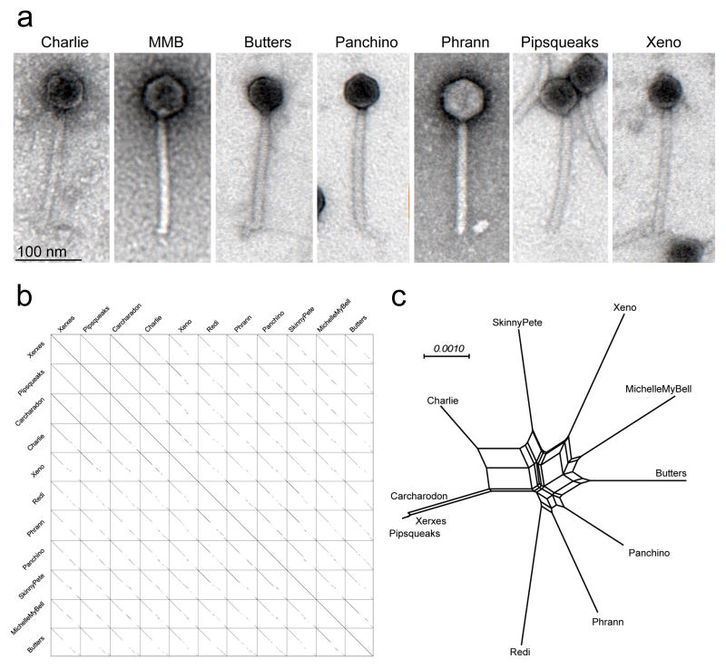
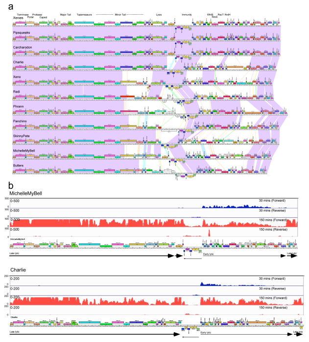
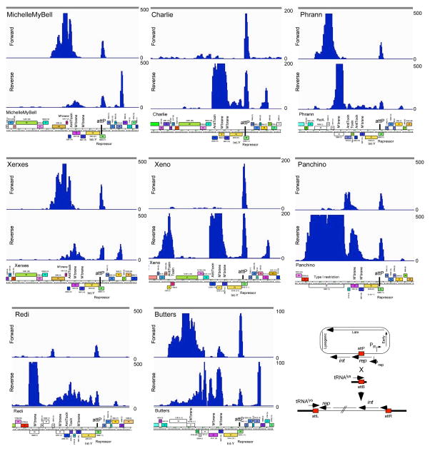
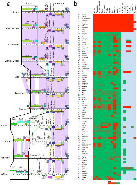
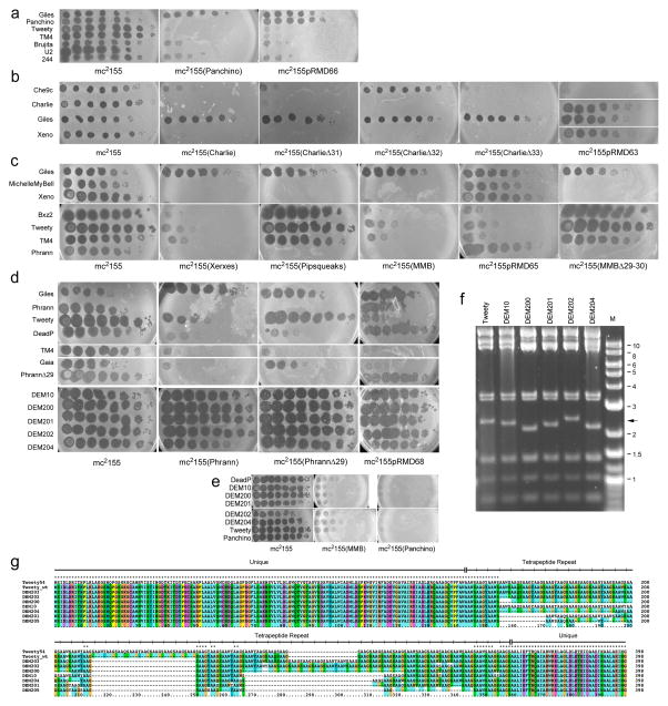
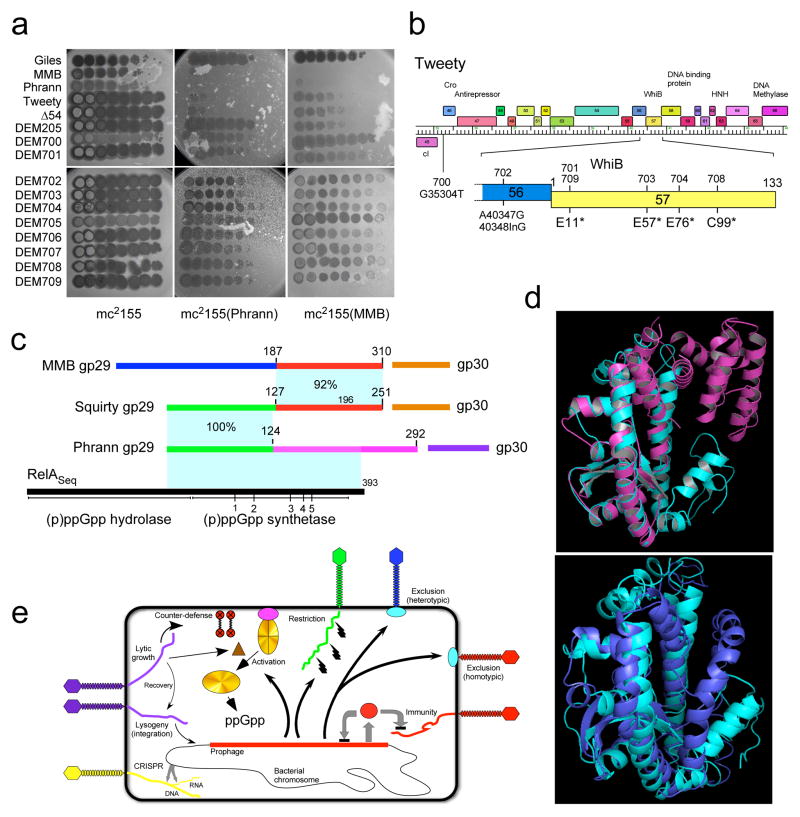
Temperate phages are common, and prophages are abundant residents of sequenced bacterial genomes. Mycobacteriophages are viruses that infect mycobacterial hosts including Mycobacterium tuberculosis and Mycobacterium smegmatis, encompass substantial genetic diversity and are commonly temperate. Characterization of ten Cluster N temperate mycobacteriophages revealed at least five distinct prophage-expressed viral defence systems that interfere with the infection of lytic and temperate phages that are either closely related (homotypic defence) or unrelated (heterotypic defence) to the prophage. Target specificity is unpredictable, ranging from a single target phage to one-third of those tested. The defence systems include a single-subunit restriction system, a heterotypic exclusion system and a predicted (p)ppGpp synthetase, which blocks lytic phage growth, promotes bacterial survival and enables efficient lysogeny. The predicted (p)ppGpp synthetase coded by the Phrann prophage defends against phage Tweety infection, but Tweety codes for a tetrapeptide repeat protein, gp54, which acts as a highly effective counter-defence system. Prophage-mediated viral defence offers an efficient mechanism for bacterial success in host-virus dynamics, and counter-defence promotes phage co-evolution.


Figures

Figure 1
Figure 1. Cluster N phage genotypes and morphotypes
(a) Electron micrographs of Cluster N mycobacteriophages. Images are representative of more than 30 particles examined from two preparations of each phage. (b) Dotplot comparison of Cluster N mycobacteriophages. A concatenated string of 10 cluster N phage genomes (as shown) was compared against itself using default parameters in Gepard. (c) Network phylogenetic analysis of Cluster N mycobacteriophages based on gene content and displayed using the Network function in Splitstree. Xerxes, Pipsqueaks, and Carcharadon have near-identical gene content, and differ by 30–50 single nucleotide polymorphisms (SNPs) and a few small insertions/deletions. Nucleotide sequence and gene content analyses do not support subclusters groupings.
Figure 2
Figure 2. Genomic organization of Cluster N mycobacteriophages
(a) The genomes of 11 Cluster N mycobacteriophages are shown with pairwise nucleotide sequence similarity displayed by spectrum-coloring between the genomes; violet is the most similar, red is the least similar above threshold BLASTN E value of 10−5. The genes are shown as colored boxes above (transcribed-rightwards) or below (transcribed leftwards) each genome, and are color-coded according to the gene phamilies they are assigned to ,; the phamily number is shown above each gene with the number of phamily members in parentheses. Maps were generated using Phamerator and the database ‘Actinobacteriophage_554’ . (b) Lytic gene expression patterns in phages MichelleMyBell and Charlie. RNAseq profiles are shown for forward and reverse DNA strands (as indicated) at 30 mins (blue) or 150 minutes (red) after infection of M. smegmatis. The numbers of reads are shown on the y-axes and genome maps are shown below.
Figure 3
Figure 3. Transcription in Cluster N lysogens of M. smegmatis
The central portions of eight Cluster N genomes are shown with their RNAseq profiles from lysogenic cultures. The numbers of reads are shown on the y-axes, and genome maps are shown below. Note that for simplicity the sequence reads are aligned to the viral rather than the prophage representation of the genomes, such that bacterial DNA sits adjacent to the left and right of attP. The cartoon at bottom right illustrates the integration system. RNAseq profiles of the entire genomes are shown in Supplementary Figures 13–15.
Figure 4
Figure 4. Cluster N prophage-mediated defense against phage infection
(a) The central parts of 11 Cluster N mycobacteriophage genomes are aligned by their immunity cassettes and putative gene functions are indicated. Genomes are displayed as described for Figure 2, but ordered such that genomes with similarities in these regions are adjacent to each other, particularly Xerxes, Carcharodon, Pipsqueaks, and MMB as one group, and Xeno, SkinnyPete and Charlie as a second group. Note that in Pipsqueaks the open reading frame corresponding to MMB 29 is interrupted by a 25 bp deletion, leaving only the 3’ end of the gene intact and presumably inactivating it. (b) Heat map of prophage-mediated viral sensitivities, reporting efficiencies of plating of mycobacteriophages on Cluster N lysogenic strains or their deletion derivatives. Individual genome names and cluster designations are shown on the left. Phages that do not have an integrase gene and are presumably lytic are shown in bold type; all others are temperate or derivatives of temperate phages. Bright green and bright red corresponds to efficiencies of platings of 1 and less than 10−4, respectively. All efficiencies of plating were determined in at least two separate experiments. Efficiencies of platings are reported in Supplementary Table 2.
Figure 5
Figure 5. Genetics of Cluster N prophage-mediated defenses
(a–e) Ten-fold serial dilutions of phage lysates were spotted onto lawns of M. smegmatis mc2155, lysogens and lysogens of mutant phage derivatives, as indicated beneath each panel. Phage names are shown to the left, including mutants (e.g. PhrannΔ29) and Defense Escape Mutant (DEM) derivatives of Tweety that overcome Phrann-mediated defense. All platings were performed in at least two separate experiments. (f) Bam HI restriction digestion of DEM DNAs shows changes in a 2.4 kbp fragment containing gene 54 (arrow). Marker (M) fragments are shown in kbp. (g) ClustalX alignment of gp54 of Tweety and DEM mutants. The wild type Tweety genome was re-sequenced and 54 contains 39 repeat units, rather than 48 in the original sequence ; repeat consensus: AAXX, where XX is either GS, GY, WS, WY, QS, or QY) flanked by unique sequences.
Figure 6
Figure 6. Mechanisms of prophage-mediated defense against viral attack
(a) Ten-fold serial dilutions of Defense Escape Mutant (DEM700-DEM709) derivatives of a TweetyΔ54 parent that overcome Phrann defense were spotted on wild type M. smegmatis (mc2155) or Phrann and MMB lysogens. All platings were performed in at least two separate experiments. (b) Tweety Δ54 DEM mutants map in early lytic genes. Genome sequencing shows DEM 700-series mutants map in Tweety early lytic genes. DEM706 and DEM707 are siblings of DEM701 and DEM702, respectively (see text and Supplementary Information for details; Supplementary Figure 22). (c–e) Relationships among phage-encoded (p)ppGpp synthetase-like proteins. (c) Phrann gp29 is a homologue of RelA/SpoT proteins with similarity to the (p)ppGpp synthetase domain of Streptococcus equimilis RelA including 5 conserved motifs (1–5). The Cluster F phage Squirty encodes a related protein (gp29) sharing the N-terminal 124 residues with Phrann gp29, but with divergent C-termini. MMB gp29 is not predicted by HHpred to be related to RelSeq, but shares its C-terminus with Squirty gp29. MMB gp30 and Squirty gp30 are closely related and are both predicted to be membrane localized. (d) I-TASSER alignment of Phrann gp29 (top, cyan) to Streptococcus equimilis RelA (PDB: 1vj7A, magenta; TM-score 0.539) and to a PHYRE2 structural prediction of Phrann gp29 (bottom, cyan). (e) Models for prophage-mediated viral defense. An integrated prophage (red line) confers defense against viral attack through numerous mechanisms, either homotypically (i.e. against the same or closely viruses) or heterotypically (against unrelated phages). Homotypic defense includes repressor-mediated immunity (repressor, red circle) and superinfection exclusion (blue circle) against itself (red phage). Heterotypic defense includes an exclusion-like system illustrated by Charlie gp32 defense against Che9c (blue phage), and restriction against many viruses (illustrated by the green phage) by Panchino gp28. Defense is also mediated by a predicted (p)ppGpp synthetase (e.g. Phrann gp29; gold circle), which we propose is kept in an inactive form (gold circle with crossed lines) by an inhibitor (purple circle), which for Squirty gp30 is membrane located. Lytic growth by specific phages activates the defense through early lytic protein, which is proposed to dissociate the (p)ppGpp synthetase from its inhibitor, enabling rapid accumulation of (p)ppGpp and growth arrest. Tweety encodes a counter-defense system (gp54) that may prevent activation of (p)ppGpp synthesis.

References

    1. Hambly E, Suttle CA. The viriosphere, diversity, and genetic exchange within phage communities. Curr Opin Microbiol. 2005;8:444–450. - PubMed
    1. Abedon ST. Phage evolution and ecology. Adv Appl Microbiol. 2009;67:1–45. - PubMed
    1. Jacobs-Sera D, et al. On the nature of mycobacteriophage diversity and host preference. Virology. 2012;434:187–201. doi: 10.1016/j.virol.2012.09.026. - DOI - PMC - PubMed
    1. Hendrix RW. Bacteriophages: evolution of the majority. Theor Popul Biol. 2002;61:471–480. - PubMed
    1. Suttle CA. Marine viruses--major players in the global ecosystem. Nat Rev Microbiol. 2007;5:801–812. doi: 10.1038/nrmicro1750. - DOI - PubMed