. 2017 Feb;24(2):184-186.
doi: 10.1038/nsmb.3352. Epub 2017 Jan 9.

Structure of the immature Zika virus at 9 Å resolution

Affiliations

Structure of the immature Zika virus at 9 Å resolution

Vidya Mangala Prasad et al. Nat Struct Mol Biol. 2017 Feb.

Abstract

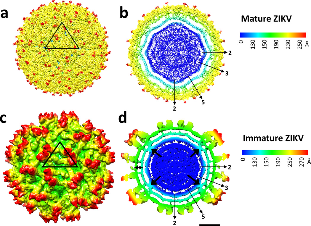
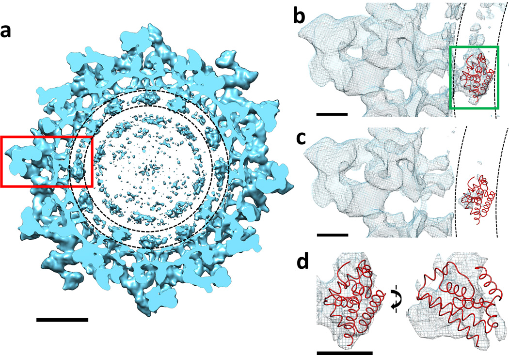
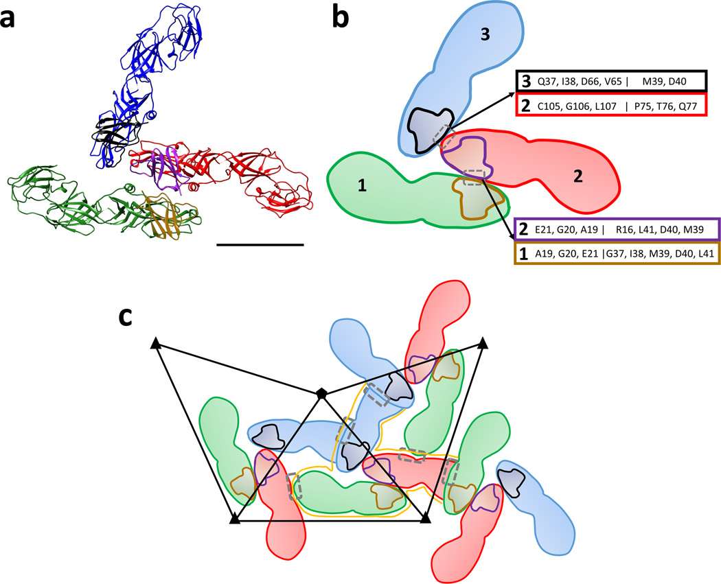
The current Zika virus (ZIKV) epidemic is characterized by severe pathogenicity in both children and adults. Sequence changes in ZIKV since its first isolation are apparent when pre-epidemic strains are compared with those causing the current epidemic. However, the residues that are responsible for ZIKV pathogenicity are largely unknown. Here we report the cryo-electron microscopy (cryo-EM) structure of the immature ZIKV at 9-Å resolution. The cryo-EM map was fitted with the crystal structures of the precursor membrane and envelope glycoproteins and was shown to be similar to the structures of other known immature flaviviruses. However, the immature ZIKV contains a partially ordered capsid protein shell that is less prominent in other immature flaviviruses. Furthermore, six amino acids near the interface between pr domains at the top of the spikes were found to be different between the pre-epidemic and epidemic ZIKV, possibly influencing the composition and structure of the resulting viruses.


Figures

Figure 1
Figure 1
Cryo-EM structure of immature Zika virus. Surface view (a and c) and cross-section (b and d) of mature and immature ZIKV colored radially. Black arrows indicate the density between the inner RNA core and the viral membrane (double-ended arrow shows the inside and outside layers of the membrane) in immature ZIKV. The asymmetric unit is given as a black triangle in panels a and c. Scale bar is 100 Å long.
Figure 2
Figure 2
Cross-section of the immature ZIKV contoured to show the inner capsid shell. (a) Cross-section of immature ZIKV projected down an icosahedral 2-fold axis. The putative location of the inner capsid shell lies between the dashed concentric circles. Scale bar is 100 Å in length. (b and c) Magnified view of the red rectangle region in panel a showing the fitted NMR structure of the DENV-2 capsid protein (PDB ID: 1R6R). Panel b is rendered at the same contour level as panel a whereas panel c is rendered at a higher contour level, showing that the capsid protein density height is only about half of that of the outer glycoproteins. (d) Magnified view of the dark green rectangle in panel b showing the fitted DENV-2 capsid protein structure along two orthogonal views. Scale bar is 25 Å long in panels b, c and d.
Figure 3
Figure 3
Interface between prM-E heterodimers. (a) Ribbon diagram of prM-E heterodimers in a trimeric spike of immature ZIKV. The prM-E heterodimers are colored in green, red and blue for the E proteins whereas the pr domains are outlined in brown, purple and black, respectively. Scale bar is 50 Å long. (b) Diagram of the prM-E arrangement in panel a using the same color coding. The three prM-E molecules are marked 1, 2 and 3. The two different interaction regions within trimeric spikes are indicated by a grey box. The molecule number and the residues involved on either side of an interaction surface are given in boxes that are colored according to the protein involved. (c) Diagrammatic representation of the trimeric spikes across adjacent asymmetric units. The interaction regions between E proteins from different spikes are indicated as grey rectangles. A single trimeric spike is outlined in orange. The asymmetric unit is outline with a black triangle.

References

    1. Schuler-Faccini L, et al. Possible association between Zika virus infection and microcephaly - Brazil, 2015. MMWR Morb. Mortal. Wkly. Rep. 2016;65:59–62. - PubMed
    1. Chan JF, Choi GK, Yip CC, Cheng VC, Yuen KY. Zika fever and congenital Zika syndrome: An unexpected emerging arboviral disease. J. Infect. 2016;72:507–524. - PMC - PubMed
    1. Lindenbach BD, Murray CL, Thiel H-J, Rice CM. Flaviviridae: The viruses and their replication. In: Knipe DM, et al., editors. Fields Virology. Lippincott Williams & Wilkins; 2013.
    1. Mukhopadhyay S, Kuhn RJ, Rossmann MG. A structural perspective of the flavivirus life cycle. Nat. Rev. Microbiol. 2005;3:13–22. - PubMed
    1. Kuhn RJ, et al. Structure of dengue virus: implications for flavivirus organization, maturation, and fusion. Cell. 2002;108:717–725. - PMC - PubMed

METHODS-ONLY REFERENCES

    1. Tang G, et al. EMAN2: an extensible image processing suite for electron microscopy. J. Struct. Biol. 2007;157:38–46. - PubMed
    1. Scheres SH. RELION: implementation of a Bayesian approach to cryo-EM structure determination. J. Struct. Biol. 2012;180:519–530. - PMC - PubMed
    1. Guo F, Jiang W. Single particle cryo-electron microscopy and 3-D reconstruction of viruses. Methods Mol. Biol. 2014;1117:401–443. - PMC - PubMed
    1. Pettersen EF, et al. UCSF Chimera--a visualization system for exploratory research and analysis. J. Comput. Chem. 2004;25:1605–1612. - PubMed
    1. Katoh K, Standley DM. MAFFT multiple sequence alignment software version 7: improvements in performance and usability. Mol. Biol. Evol. 2013;30:772–780. - PMC - PubMed

Publication types

Substances